Pancreas
The ultrasonographic examination of the pancreas is highly operator dependent and an excellent knowledge of anatomy, including vascular anatomy as well as experience with ultrasonography is required to locate the pancreas and interpret its appearance.
High resolution transducers are required to image the pancreas in normal animals. Excessive gas in the gastrointestinal tract as well as deep thoracic conformation may inhibit visualization of the entire pancreas. Obese animals pose an additional barrier, even for experienced ultrasonographers. Furthermore, animals with pancreatic disease often have abdominal pain and resent transducer pressure in the region of the pancreas, and analgesia may be necessary in order to perform a thorough examination. Another ultrasound tool for examination of the pancreas in either obese or painful animals is echoendoscopy. Using an echoendoscope, a high frequency transducer can be inserted into the stomach and the pancreas is imaged transgastrically.59 Obesity and air pose fewer problems for echoendoscopic imaging compared to conventional transabdominal ultrasound. The author has been able to consistently examine the pancreas using this technique in both dogs and cats.1.3.8.1 Pancreatitis
While ultrasound is generally considered a valuable tool for the diagnosis of canine pancreatitis, its sensitivity in cats with pancreatitis is variable.60 In patients with acute pancreatitis the pancreatic parenchyma becomes hypoechoic and enlarged. The surrounding mesentery of the pancreas is often diffusely hyperechoic and poorly circumscribed. However, these changes may be subtle in cats. When the mesenterium is inflamed and the bowel loops are distended with gas, the pancreatic tissue itself may be difficult to identify ultrasonographically (Figure 1.40). Mild to moderate accumulations of free fluid may be detected in the cranial abdomen.
Together with a hyperechogenic mesentery this may indicate the presence of focal peritonitis. In such cases, the small intestinal loops and especially the duodenum may be dilated due to functional ileus. This may contribute to lack of visualization of the pancreas due to fluid and gas accumulation. In cats, multiple hypo- echoic round foci of a few millimeters in diameter may be recognized if high frequency (greater than 7.5 MHz) transducers are used. These findings may present either nodular hyperplasia or dilated pancreatic ducts.61Figure 1.41:
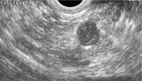
Canine insulinoma. Echoendoscopic image of the body of the pancreas in a 7-year-old, female Bearded Collie with hypoglycemia. The pancreas was found to be normal in the transabdominal ultrasound examination. An 8.6-mm diameter, round, and hypoechoic nodule was identified echoendoscopically. The nodule was removed surgically and was diagnosed histologically as an insulinoma.
The ultrasonographic appearance of chronic pancreatitis in dogs and cats has not been well described. Recurrent episodes of pancreatitis may lead to chronic changes that can be identified ultrasonographically and are mainly due to fibrosis. The pancreas may be of normal size or enlarged with a heterogenous appearance. Mineralizations may be present and may lead to acoustic shadowing.
Cavities in the pancreatic parenchyma are typically either due to abscesses or pseudocysts and appear as anechoic or hypo- echoic cavities, possibly with a thickened wall. Fine-needle aspiration for cytological analysis may be useful to differentiate them.
1.3.8.2 Pancreatic neoplasia
Pancreatic neoplasia is much less common than pancreatitis in both dogs and cats.62 Neuroendocrine tumors are the most common pancreatic neoplasia, followed by adenocarcinoma and metastatic tumors. Differentiation of pancreatitis and pancreatic neoplasias with ultrasound is not always easy due to the overlapping nature of their appearance.62 Lymphadenomegaly can occur with both and surrounding tissues are often similarly altered.
Fine-needle aspiration or true-cut, laparoscopic, or surgical biopsy is often needed for conclusive differentiation. Involvement of multiple organs can occur in both severe suppurative pancreatitis or primary liver, pancreatic, or bile duct neoplasia.62 Neoplasia of any one of these structures may invade adjacent organs and simulate inflammatory or granulomatous disease. In such cases, biopsy of both the liver and pancreas is warranted. Diffuse infiltrative disease of the pancreas, liver, and other organs such as the stomach, duodenum, spleen, and lymph nodes may be seen in suppurative, granulomatous, as well as neoplastic disorders.Depending on the size of a pancreatic nodule, the amount of gastrointestinal gas, and thoracic conformation of the patient, pancreatic neoplasms such as insulinomas and adenocarcinomas may be difficult to detect ultrasonographically. Endosonography of the pancreas via a transgastric approach may allow better assessment of the entire pancreas in dogs (Figure 1.41).59 The author has been able to diagnose a number of insulinomas echoendoscopically that could not be visualized with conventional transabdominal ultrasound. Recently, CT and nuclear scintigraphy using radiolabeled leucocytes has been evaluated for assessing the canine and feline pancreas.11,63 The MRI appearance of the normal feline pancreas has also been reported, but reports of its clinical use are lacking.64 Such modalities may have future potential for diagnosing pancreatic disease but more clinical data is needed.
Key Facts
■ Radiography and ultrasonography are two of the most valuable diagnostic tools for diagnosing gastrointestinal disease in dogs and cats.
■ Thoracic radiography should be performed in every patient with regurgitation in order to assess the esophagus and rule out aspiration pneumonia. However, dynamic contrast studies are often necessary to diagnose the cause of dysphagia.
■ High quality radiographs of the abdomen in two orthogonal planes should be performed in patients presented for vomiting and are important for ruling out obstructive ileus. Abdominal ultrasound and /or barium studies may be indicated when radiography does not allow definitive ruling out of an obstructive lesion.
■ Ultrasound is indicated in dogs and cats presenting with icterus. Hepatic vs. post-hepatic causes can be investigated and ultrasound- guided biopsies of the liver or pancreas can be performed.
■ Abdominal ultrasound is clinically useful for the diagnosis of pancreatitis in both dogs and cats. The examination should include assessment of the liver, gall bladder, mesentery, and regional lymph nodes in order to rule out multi-organ involvement.