Periparturient Problems
A variety of serious complications can interfere with normal parturition. Frequent close observation of the doe is the best insurance that difficulties will be recognized in time to save doe and kids.
Pregnancy Toxemia
Does that are carrying multiple fetuses may become anorectic and ketonuric in the last four to six weeks of gestation if feed intake does not match the metabolic needs of both the dam and the fetuses. Possible sequelae include dystocia and stillbirth or even death of the dam. A full discussion of pregnancy toxemia is included in Chapter 19. It is very important to check late-pregnant goats for ketone- mia or ketonuria if they show any signs of ill health. Treatment of the ketotic state is equally important whether the condition is primary (caused by improper nutrition) or secondary to some other disease or misadventure.
Prolapsed Vagina
Prepartum eversion of part or all of the vagina leads to discomfort and straining by the doe. This results in eversion of more tissue (Figure 13.9). Infection or laceration of the wall of the vagina and loss of the cervical seal (with subsequent entry of infection into the uterus) commonly occur. If the bladder becomes incorporated into the prolapse, kinking of the urethra impedes urination while enlargement of the bladder provokes even more straining. Rarely, the vaginal wall may rupture, leading to evisceration of intestines or even prolapse of the pregnant uterus through the tear (Brunsden et al. 2020).
Causes of vaginal prolapse are not well understood in goats, but might include heredity, increased intraabdominal pressure from a large litter or indigestible roughage diet, excess estrogen in the forage, lack of exercise, or previous prolapse (Braun 2007). Unfortunately, failure of cervical dilation often leads to dystocia when labor begins after a vaginal prolapse.
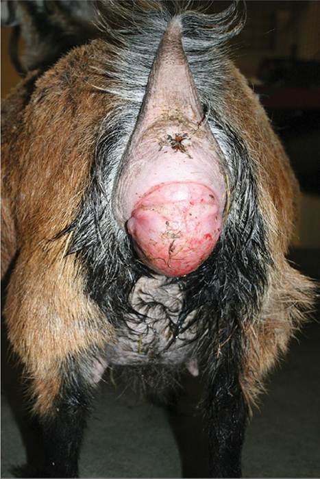
Figure 13.9 Prepartum prolapse of the vagina. The cervix of this pygmy doe failed to dilate when she went into labor, necessitating a cesarean section. Source: Courtesy of Dr. M.C. Smith.
The anatomy, including innervation, of the pelvic outlet of the doe has been reported in detail (Hartman 1975).
Management of Mild Prolapses
A mild prolapse is one that is visible when the doe is lying down but disappears when the animal stands up. Sometimes this condition can be handled by restraining the doe full time in such a way that her front end is lower than her hind end whenever she lies down. Possibilities include tying the doe in a corner where an excavation big enough for her frontquarters has been made in the bedding pack, or confining the doe to a narrow pen such as a metabolic cage and then raising the posterior end of the platform on which she stands and sleeps.
Retainers and Trusses
A prolapse that does not correct itself when the doe stands up should be cleansed with a non-irritating soap, coated with an emollient or oily antibiotic preparation that includes a topical anesthetic, and manually replaced. A caudal epidural block (see Chapter 17) is helpful to limit straining during the procedure. If the prolapse appears to be too large to replace, lift up from below to unkink the urethra and permit emptying of the trapped bladder. Then some means must be found to keep the vagina in its normal position until parturition brings a decrease in the dimensions of the vagina and the estrogen-induced relaxation of surrounding tissues. Parturition is usually not possible until the vagina has been replaced. A paddle-shaped plastic bearing retainer is frequently used to treat vaginal prolapses in sheep. The same device can be used in goats, but a way must be found to attach it to the doe. In small breeds a syringe case can be used in place of a paddle. Where a fleece is lacking, a length of umbilical tape can be glued to the doe's hair on each side of the hindquarters, near the point of the hip, and the two ends of the retainer are then tied to these tapes.
Alternatively, a loop of suture can be placed in the skin on each side for attachment of the umbilical tapes. Presence of the retainer does not prevent kidding.A rope truss that puts external pressure on each side of the vulva is also effective in sheep and Angora goats, but it is more difficult to keep the truss properly adjusted on the slippery haircoat of other breeds of goats. Still, the truss has the distinct advantages that it does not provoke straining by the doe and introduces no danger of infection. The center of a long piece of clothesline is placed over the doe's neck. The ends are then crossed under the chest (between the front legs) and crossed again over the back. They are then passed between thigh and udder on each side, continuing up along the vulva on the same side and forward around the cross in the middle of the back, to be secured to the rope over the neck. Short pieces of rope are used to connect the two lines above and below the base of the tail and just below the vulva. Alternatively, supporting pressure can be applied to the vulva by means of a stiff rubber, leather, or aluminum rod truss of triangular shape, with loops at the two upper lateral and single ventral corners simplifying attachment to the rope harness (Babin et al. 1981). Commercial prolapse harnesses made of webbing are also available, and a pony halter placed on the frontquarters of the goat may help to hold the truss in place on a smallbreed doe.
Vulvar Sutures
Other methods of retaining a prolapsed vagina include several deep mattress or cruciate sutures across the vulva and a buried pursestring (Rahim and Arthur 1982). Local anesthesia by means of a caudal epidural is imperative (see Chapter 17). The doe must be watched very carefully, because the sutures prevent parturition and cause tissue infection. A less invasive technique involves bootlacing across the vulva with a soft strip of cloth or gauze, using small loops of umbilical tape that are placed in rows quite lateral to each side of the vulva.
A single anchoring loop between vulva and anus serves to prevent the lacing from slipping ventrally. The owner can loosen the lacing and check on the progress of parturition without causing further tissue damage or discomfort to the doe.Milk Fever and Uterine Inertia
Clinical signs of hypocalcemia (inappetence, unsteady gait, weakened uterine contractions, eventual recumbency) are occasionally noted in periparturient goats. Also, a mild hypocalcemia might contribute to prolongation of first- stage labor. If milk fever is suspected, calcium borogluco- nate (60-100 mL of a 20-25% solution) can be given very slowly intravenously or subcutaneously, split into four sites. Products with phosphorus and dextrose should be avoided subcutaneously, because they cause painful abscess formation. Intravenous infusion should be avoided if the animal appears to be toxic. Other explanations for the illness, such as indigestion, pregnancy toxemia, ruptured uterus or uterine artery, metritis, and mastitis, should be ruled out. Milk fever is discussed in detail in Chapter 19.
Failure of Cervical Dilation
“Ringwomb,” or incomplete dilation of the cervix, is observed occasionally in goats, as in sheep. In one study in Iraq, ringwomb accounted for 24% of 136 goats presented to a referral clinic for dystocia (Majeed and Taha 1989). In another report, ringwomb was diagnosed in 33% of 284 goats presented in dystocia to a teaching hospital in Saudi Arabia (Ali 2011). The etiology remains unexplained in both species. Primiparous does may be predisposed to the condition. Undoubtedly, some cases diagnosed as incomplete dilation merely represent too early intervention. Others are accompanied by pregnancy toxemia, uterine inertia, or fetal malpresentation (Rahim and Arthur 1982) or uterine torsion (Ali 2011). Sometimes the cervix is actually closing again after failure of parturition to occur at the appropriate time. When the cervix only opens enough for a small sack of placenta to pass and the fetuses are dead or premature, it seems likely that inadequate hormonal preparation for parturition may have been involved, as with diseases involving the placenta.
Toxoplasmosis is one disease that has been documented to affect hormone production by the placenta (Engeland et al. 1996).Possible treatments include careful manual stretching of the cervix (success in 2 of 16 cases; Ghosh et al. 1992), administration of calcium and/or estrogen and waiting a few hours, and cesarean section. Prostaglandin F2 alpha (7.5 mg intramuscularly) has been proposed as an effective treatment, as judged by delivery of kids within four hours (Majeed and Taha 1989). Cloprostenol (500 μg intramuscularly) has also been reported to be effective, though the time to delivery was reported to be 39-51 hours (Ghosh et al. 1992). The possibility that the final preparturient surge of prostaglandin is absent in animals with ringwomb needs to be investigated. It is tempting to ascribe a response to prostaglandin to induction of relaxin, a glycoprotein hormone that softens the cervix in some species (Senger 2005), but relaxin has not yet been adequately studied in the doe. If a true ringwomb is present, manual dilation can easily rupture the cervix or uterus (Engum and Lyngset 1970). Oxytocin also should be avoided. If placenta is visible and other techniques fail, surgery should not be delayed longer than a few hours. However, when the kids were already dead, Ali (2011) reported success with administration of 15 mg prostaglandin F2 alpha and waiting for full dilation of the cervix to occur in an average of 42 hours in 35 of 51 does. Those that failed to dilate were then subjected to a cesarean section.
Abnormal Fetal Presentation, Position, or Posture
Both anterior presentation (with head and forelimbs extended) and posterior presentation (with hindlimbs extended) are considered to be normal in goats. However, posterior presentation has been reported in only 3-9% of kids, is even rarer in single kids, and appears to predispose to dystocia (Engum and Lyngset 1970). Problems occur when multiple kids become tangled in the birth canal or the head or limbs are retained.
Careful attention is necessary to distinguish front feet from hind feet and to verify that traction is being applied to the extremities of only one fetus at a time. Normal deliveries and various dystocias in goats have been well illustrated in a French publication (Babin et al. 1981).Some breeders report increased frequency of dystocia in Pygmy goats and suggest that selection in the show ring for a very short and compact body type is to blame (Brown 1988). Apparently there is not enough room in the doe's abdomen for the kid to present itself properly. A radiographic study of dystocia in 30 West African Dwarf goats in Nigeria, with no show ring selection, demonstrated that abnormal fetal posture was more common than a small maternal pelvis as a cause of dystocia (Kene 1991). Pygmy goats were overrepresented in one study of goats undergoing cesarean section in the United States (Brounts et al. 2004), but this might be partly due to inadequate space in the goat's pelvis for manual correction of a dystocia.
General Guidelines for Mutation and Delivery
Wild goats are said to stand with their forequarters downhill from the hindquarters to give fetuses the opportunity to rearrange themselves. This gravitational advantage should be remembered when parturition is assisted. If it is necessary to retrieve the head or a limb of a fetus in a recumbent doe, this is easiest if the dam is positioned so that the retained extremity is uppermost. Delivery is usually possible with either one forelimb or one hindlimb retained. If there is adequate room for manipulations, the second limb can be properly positioned before extraction begins. If the head is out, it is sometimes possible to deliver the normalsized kid by simple traction, without retrieving either forelimb. If the uterus is contracted around the fetus subsequent to prolonged dystocia, intramuscular epinephrine (1 mL of a 1 mg/mL solution to a dairy or meat breed doe) greatly facilitates safe manipulations by briefly relaxing the uterus.
When the kid is properly positioned, it is pulled out and downward in an arc. If a dystocia occurs when head and two forefeet are presenting, the difficulty may be a simple elbow lock; pulling firmly on one limb at a time extends the elbow joint so that the tip of the nose is positioned closer to the carpus rather than directly over the hooves. It is also possible that parts of more than one fetus are represented or that a relative fetal oversize is present.
Deviation of the Head
One should never try to extract a kid in anterior presentation unless the head and neck have been either properly positioned or amputated. Rupture of the uterus is very likely to occur and may already be present before human intervention when the fetal head is retained. A lamb puller or lamb and pig puller (available from various supply houses) makes an excellent head snare. The head typically deviates to the side of the fetus, but occasionally drops down between the front limbs. It is usually necessary to repel the fetus by pushing the front limbs inward or elevating the hindquarters of the doe to have space to straighten out the head. Epinephrine, as described above, may also be helpful.
Retention of Forelimbs
Sometimes the head of a kid is delivered, but then progress stops because of retention of both forelimbs. Assistance should be provided at once; the neck is followed to a shoulder and then the corresponding limb is retrieved. It may be necessary to repel the head to have room for passing a hand as far as the shoulders. Care must be taken not to push the head around to one side, and a head snare might be placed before repelling the head.
If the head of a live kid with retained forelimbs passes beyond the lips of the vulva, compression of the jugular veins results in edema of the head. The head swells to the extent that repulsion becomes impossible yet there is no room to reach inside and straighten a leg. Extensive lubrication may permit delivery of a small kid in this position without additional manipulation. If the kid is dead, there should be no hesitation about amputating the head at the atlanto-occipital joint with a scalpel blade to provide more room for mutation.
Multiple Fetuses or Transverse or Malformed Fetus
When more than one kid is presented simultaneously, it is important to accurately identify the extremities in the birth canal and to trace them back to the body of the fetus. In general, it is preferable to first deliver a kid whose head is in the birth canal rather than risk creating a more difficult dystocia by repelling the head. Otherwise, a kid in posterior presentation is easier to deliver first. If a kid is transversely positioned, it should be moved (with the finger tips) into a posterior, breech position. From this point, retrieval of at least one hindlimb usually permits delivery. Epinephrine to relax the uterus, as described above, may be helpful.
A schistosomus reflexus fetus (Buergelt 1997; Gutierrez et al. 1999; numerous other individual case reports) could cause dystocia that resembled the simultaneous presentation of twins. If correctly diagnosed by noting abdominal organs outside the fetal body, the dystocia can be relieved by a single cut with a regular fetotome.
Controlling Straining
An epidural injection of lidocaine (with or without xylazine) can be used to decrease straining, as discussed in Chapter 17. The beta-adrenergic compound clenb- uterol (Ventipulmin®, Boehringer Ingelheim) is a.va.ila.- ble in some countries as a tocolytic drug to temporarily cause myometrial relaxation. Initial favorable studies in cattle suggest that the drug might also be useful during correction of fetal malposition or exteriorization of the uterus during cesarean section in goats. A possible dose to try is 0.8 μg∕kg intramuscularly (Menard and Diaz 1987). The use of this drug in food animals is IiaTidden in the United States, but epinephrine may be substituted (see above).
Relative Fetal Oversize and Fetotomy
Multiple kids are rarely too large for the birth canal, unless the doe is severely stunted or has suffered some injury such as ankylosis of a fractured tail (Engum and Lyngset 1970). On the other hand, a single large kid may be too large for a small first freshener. Owners rarely have the luxury of breeding doelings to a buck of known kidding ease. Addition of lubricant to the birth canal may assist delivery of a large kid, but often the fetus is emphysematous, malformed, or too large to be extracted intact.
Fetal hydrops (anasarca, dropsy) is a specific cause of fetal oversize that warrants discussion. Massive accumulation of fluid in fetal tissues and body cavities may make extraction impossible unless incisions are first made with a finger knife to drain the fluid. Accumulation of fluid within the placenta may be so great that abnormal distension of the dam's abdomen occurs, and the subsequent interference with circulation, respiration, and locomotion necessitates a cesarean section or induction of parturition to save the dam. Anasarcous kids may be co-twin to normal, viable kids, and the same doe may produce another affected kid in a subsequent gestation (Elze and Muller 1960; Walser 1963; Sarangom et al. 2020). In one herd, 11 kids with hydrops were produced, all sired by a single buck. The condition may be inherited as a simple recessive character (Ricordeau 1981). Hypothyroidism is another proposed cause (Kumar et al. 1989). Wesselsbron disease (Chapter 11) is also reported to cause hydrops, at least in sheep.
Unless the kid is alive and its value warrants the expense of a cesarean section, consideration should be given to a subcutaneous fetotomy. This procedure is relatively safe for the doe because instruments are not used in the birth canal and the fetal skin protects the friable maternal tissues. An initial skin incision is made with a scalpel just above and all the way around the carpus, and is then extended as far as possible up the medial side of the leg. The skin is then pushed proximally and the front limb is torn loose from the thorax and extracted, an easy process if the fetus is autolyzed. This procedure is repeated with the other front limb. The thorax can be compressed by sliding a hand beneath the kid's skin and manually fracturing the ribs. Rotation of the head and trunk causes the vertebral column to fall apart in the lumbar region. The hindlimbs can then be pulled out. A complete subcutaneous fetotomy is rarely necessary (Engum and Lyngset 1970).
Special tools have been described for fetotomy in small ruminants. A long-handled nipper with blunt-tipped jaws bent upward has been used to amputate a front or back limb or the head. Fetal pieces are then extracted with the aid of small tongs (Deckwer 1951).
Hydrops of the Uterus
Rarely, distention of the pregnant uterus with excess amniotic or allantoic fluid occurs. Extreme abdominal distension, discomfort, and difficulty rising may be noted. Presence of caruncles and fetus on ultrasound distinguishes hydrops from hydrometra. Pregnancy toxemia (see Chapter 19) may be concomitant or incorrectly suspected. The normal allantoic fluid volume in sheep and goats has been reported to be between 0.5 and 1.5 L, while one doe with hydrallantois had approximately 12 L of fluid at the time of successful treatment by cesarean section (Morin et al. 1994). Hormonal induction of parturition permits slower release of the accumulated fluid, but the delivery is also slow and probably requires assistance because of weakness of the myometrium (Jones and Fecteau 1995; Sreejith et al. 2009). It has been proposed that ram ? doe hybrid pregnancies are predisposed to the development of hydrops of the uterus (Kelk et al. 1997).
Uterine Torsion
Torsion of the pregnant uterus is occasionally observed in goats (Wyssmann 1945) as in sheep (Smith and Ross 1985). In one report from Kerala, India, torsion was identified as the cause of dystocia in 22 of 53 goats treated by cesarean section during a five-year period (Philip 1985). The condition is more likely to occur with a single fetus than with bicornual twins. The torsion may involve the vagina, but it also may involve only the cervix or body of the uterus. Thus, when a doe is in dystocia, as evidenced by ineffective straining, it may not be possible to distinguish uterine torsion from incomplete dilation of the cervix. Early cesarean section permits differentiation and correction of both of these conditions. Ultrasound examination may demonstrate marked thickening and edema of the uterine wall near the site of torsion (Wehrend et al. 2002).
If the torsion is recognized at the time of examination for dystocia, correction may be attempted by rolling the doe while weight is applied to the abdomen to prevent rotation of the fetus (Sathiamoorthy and Kathirchelvan 2005). Thus, if the torsion palpable in vagina or cervix is to the left, the doe is placed in left lateral recumbency and rolled over her back to the other side, while the uterus is prevented from turning by hand pressure or a weighted plank (Biswal et al. 2015) held across the flank. Suspending the doe by her hindlimbs and shaking the doe or manipulating the uterus through the body wall is an older, but sometimes successful approach to repositioning the uterus.
Cesarean Section
When pregnancy toxemia or other misadventure makes immediate termination of pregnancy desirable or when a dystocia cannot otherwise be resolved without sacrificing the life of a kid, surgical delivery of the fetuses is necessary. Ultrasound can be used to assess fetal viability, as immediate slaughter might be the economically correct decision if the fetuses are dead. Research or disease eradication programs requiring gnotobiotic kids may involve elective cesarean section. Because many goats shed the Q fever organism (C. burnetii) in birth fluids, all people assisting with the surgery or aftercare of fetuses should wear gloves and tightly fitting masks.
Anesthesia and Surgical Approach
Either general or local anesthesia may be chosen (Benson 1986). Suitable general anesthetic agents (halothane, isoflurane) are rapidly cleared after surgery so that there is minimal respiratory depression of the kids or interference with maternal behavior. Xylazine tranquilization should be kept to a minimum or avoided altogether, unless reversal agents (yohimbine or tolazoline) are available. Intramuscular xylazine at 0.2 mg/kg causes hypoxemia, respiratory acidosis, uterine contractions, and decreased uterine blood flow in the pregnant goat (Sakamoto et al. 1996). Tranquilization with diazepam is far preferable to xylazine. An intravenous diazepam dose of 5 mg per goat has been suggested for standing surgery (Snyder 2007). When the doe is toxic or exhausted, local anesthesia (see Chapter 17) alone is adequate. Intravenous fluid therapy should be begun on these patients before surgery commences.
The doe may be tied in right lateral recumbency, with a towel covering the eyes. A vertical incision is made halfway down the left paralumbar fossa. Care should be taken to enter the abdominal cavity rather than the perirenal retroperitoneal space. A ventral midline incision along the linea alba starting at the base of the udder and extending crani- ally 20 cm is an alternative that requires less suturing to close, but dorsal recumbency increases pressure on major blood vessels and the danger of regurgitation. Using a cuffed endotracheal tube and positive pressure ventilation overcome these disadvantages. A ventral abdominal paramedian approach, between the linea alba and the subcutaneous abdominal vein, is yet another option (Tibary et al. 2017).
Surgical Techniques
When the abdominal cavity has been opened, the greater omentum should be retracted cranially and the abdomen explored to determine the number and location of fetuses. A gravid horn is then exposed and packed off with moistened towels. A single incision along the dorsal curvature of one horn usually permits removal of all fetuses from one site (Wallace 1982). Placentomes should be avoided. Extremities (e.g., hind feet) are grasped and the first kid is gently delivered from the abdomen. Additional kids are milked to the same incision. The umbilical cord is separated by grasping it firmly close to the kid's abdominal wall with one hand and tugging firmly with the other hand, which grasps the cord several inches distally. If the cord fails to break, it should be clamped with Carmalt forceps and ligated. The placenta is only removed if it is no longer attached to caruncles. After the surgeon is sure that all fetuses have been removed, the uterus is closed with one or two layers of inverting 0 or 1 chromic catgut or synthetic absorbable sutures. After the surface of the uterus has been lavaged, closure of the abdominal incision is routine (Tibary et al. 2017).
If the doe has a problem that prevents vaginal delivery on subsequent pregnancies but it is to be kept as a pet, ovariectomy should be considered before abdominal closure, as the surest way to prevent rebreeding.
Aftercare
Aftercare of kids is as described for vaginal deliveries. Ideally, an assistant should attend to the kids, because their body temperature can drop dangerously low during the time it takes to finish the surgery. If the doe is to raise the kids, she should be permitted to mother them as soon as possible. Systemic antibiotics (penicillin, ceftiofur, or tetracycline) and tetanus prophylaxis are generally administered to the dam and ideally given before surgery commences. Oxytocin (less than five units) should not be given until suturing of the uterus is completed. Research conducted in ewes suggests that oxytocin doses exceeding 3 or 4 IU intramuscularly cause a prolonged and undesirable spastic contraction of the periparturient uterus (Marnet et al. 1985), but higher (empirical) doses have been recommended in small ruminants. Analgesics such as flunixin meglumine (1.1 mg/kg intravenously) may be given if needed to control postoperative pain (Vivrette 1986). Oral meloxicam at 1 mg/ kg might be administered for several additional days.
The prognosis for life and fertility of the doe is good if the surgery was elective (Brounts et al. 2004), but must be guarded if the fetus was macerated or the doe was seriously ill, as with pregnancy toxemia, before surgery.
Prolapsed Uterus
Eversion of the uterus (typically one horn) occurs occasionally after parturition, but is apparently less common when goats are loose-housed than when they are confined to individual stalls (Engum and Lyngset 1970). The prognosis is usually good, even if the prolapse has been present for 24 hours. The first step is a caudal epidural block, as described in Chapter 17. The uterus should be washed with copious water and a mild disinfectant. The hindquarters are then elevated with the hindlimbs spread apart; placing the back end of the goat over a hay bale of bag of shavings works well. Working through a towel while replacing the uterus protects against accidental perforation. When the uterus is back through the cervix, manipulation with fingers held together is continued until the very tip of the uterine horn is replaced. Warm water with a mild disinfectant can be pumped into the uterus to ensure complete replacement of the horns and stimulate uterine contraction. Alternatively, an antibiotic powder is placed in the uterine cavity and oxytocin (perhaps 5 IU) is given to contract the uterus. Systemic antibiotics may be given for several days. Tetanus prophylaxis should not be neglected. Some authors recommend suturing the vulva, but this should not be necessary if replacement is complete and the doe can get up easily. A truss could be used for security and would allow recognition of a repeat prolapse.
If the prolapse is older than 36 hours or is extensively damaged, amputation of the uterus is required. First verify that the bladder and intestines are not trapped within the prolapse. This can be done by inspection after carefully incising through the uterine wall. Ideally, large vessels are visualized and ligated (Miesner and Anderson 2008). Tight transfixing ligatures of the prolapse with heavy absorbable suture are required, and a stay suture should be placed ahead of the ligature, in case a ligature should slip during amputation and retrieval of the stump should be required (Engum and Lyngset 1970).
Uterine Artery Rupture
Though this condition is not well described in the literature, rupture of the uterine artery occurs occasionally in the postpartum goat. It is conceivable that some does merely experience hematoma formation in the broad ligament, but a problem is usually only diagnosed if fatal hemorrhage occurs. The doe may be found dead or dying; cold skin, muscle tremors, and bloat in the hypovolemic animal may wrongly suggest a primary diagnosis of hypocalcemia. Death may occur before the packed cell volume has time to fall. The author has seen goats that bled to death as long as four days post partum.
Retained Placenta and Metritis
The placenta is normally passed within three or four hours. If the placenta has not been expelled within 12 hours after delivery of the last kid, it is then considered to be retained (Franklin 1986). Retained placenta may occur in an infectious abortion (or birth of a live kid from an infected doe) caused by diseases such as brucellosis, toxoplasmosis (Calamel and Giauffret 1975), and chlamydiosis. Practitioners have associated a dietary deficiency of selenium with herd problems of retained placenta in goats, and recommended injectable and oral selenium supplementation for prevention (Cochran 1980). Some delay the injection until 10 days before the due date, in light of label warnings against the use in pregnant animals, reasoning that kids might still be viable if treatment induced early parturition. Cesarean section is another risk factor for retained placenta (Brounts et al. 2004).
Metritis associated with retained placenta, retained kids, or trauma and infection of the uterus in the course of dystocia may lead to signs of systemic illness shortly after parturition. Fever, depression, anorexia, and a malodorous vaginal discharge are typical. The occurrence of a retained placenta may be difficult to confirm without a vaginal examination, because goats commonly eat all or part of the placenta. In one study of 60 normal parturitions, the placentas were eaten by 38% of the does (Lickliter 1984/85). Transabdominal ultrasound during the first week, at least in sheep, permits identification of hyperechoic placenta in fluid in the uterine lumen, attached to hypoechoic caruncles (Hauser and Bostedt 2002). Peritoneal fluid analysis may suggest peritonitis if a severe metritis or uterine tear is present. If a vaginal examination is performed, intrauterine antibiotics are commonly administered, but manual removal of the placenta should not be attempted. Instead, oxytocin may be administered (5 IU subcutaneously or intramuscularly several times a day) and systemic antibiotics are recommended to prevent development of a lifethreatening toxemia or septicemia. It is the author's (MCS) personal preference to give penicillin prophylactically, but to use oxytetracycline if the goat is already systemically ill. Tetanus prophylaxis should be ensured.
Postparturient or genital gas gangrene caused by infection of the uterus with clostridial organisms is common in Angora goats in South Africa that are penned for kidding (Van Tonder 1975). Signs usually develop 6-24 hours after kidding. Metritis is accompanied by septicemia and death occurs in untreated animals in 12-72 hours.
Pyometra appears to be far less common in goats than is mucometra or hydrometra associated with false pregnancy (Hesselink and Taverne 1994). Previous dystocia with cervical damage may predispose to pyometra (Haibel 1986a), as does the presence of parts of a macerated fetus (Lyngset 1968d). The contents of the uterus are more uniformly echogenic when examined by real-time ultrasound than is the fluid in a hydrometra. Fetal bones are sometimes found embedded in the wall of the reproductive tract at necropsy. Treatment is with prostaglandin, systemic antibiotics, and possibly surgical emptying of the uterus or flushing with an iodine solution or hypertonic saline administered via a pipette.
VuLvovaginitis
Vaginal discharge (other than clear or cloudy heat mucus and lochia) or lesions of the vaginal mucosa in the absence of uterine or urinary tract disease warrant a speculum examination of the vagina. Traumatic lesions and retained vaginal pessaries should be ruled out before investigating the possibility of specific infections localized to the caudal portions of the reproductive tract. Ectopic mammary gland (see Chapter 3) should not be mistaken for inflammatory disease of the vulva.
Caprine Herpesvirus
Postbreeding genital disease (papules, vesicles, and pustules coalescing to form ulcers) associated with caprine herpesvirus-1 has been reported in does in New Zealand (Horner et al. 1982), Australia (Grewal and Wells 1986), and Italy (Camero et al. 2015. Lesions healed in four to eight days and does developed neutralizing antibody. Vaginal lesions have also been produced by experimental infection and reactivated by estrus or corticosteroids (Tempesta et al. 2002). There is no apparent interference with fertility.
Infection can be confirmed by viral isolation from vaginal swabs or by rising serum neutralization titers. The condition is discussed in Chapter 12.
MycopLasma
A granular vulvovaginitis has been observed in spontaneous cases and reproduced experimentally by inoculation of scarified vulvar mucosa with Mycoplasma agalactiae (Singh et al. 1974, 1975). Spontaneously affected goats have a mucopurulent vaginal discharge with multiple, yellowish-white, pinhead-sized elevations, especially near the clitoris. Occasionally the granules are red and ulcerated. Histologically, findings include perivascular cuffing, accumulation of lymphocytes and plasma cells in the lamina propria, and lymphoid follicles in the vulvovaginal region. Numerous other mycoplasmas, including Mycoplasma bovigenitalium and Acholeplasma Iaidlawii, have been isolated from Nigerian goats with clinical vulvovaginitis (Chima et al. 1986). A. laidlawii is found on all mucosal membranes (Rosendal 1994) and is of questionable pathogenicity, but experimentally has been reported to cause lymphocytic vulvitis and vaginitis in goats (Gupta et al. 1990).
There is no published evidence that this condition interferes with fertility or that its occurrence or duration can be limited by treatment. To avoid additional spread, however, it seems prudent to avoid natural mating until clinical signs disappear.
Leiomyofibromatosis
A prolonged vaginal discharge (purulent or serosan- guinous) has been reported from a Saanen goat with multiple firm nodular polyps in the vaginal wall. Based on histology, a diagnosis of vaginal leiomyofibromatosis was made and an association with follicular ovarian cysts was proposed (Haibel et al. 1990). One of the authors (MCS) is aware of a similar case in a goat with bilateral granulosa cell tumors. Because the diagnosis was only considered retrospectively after review of the first report, it seems likely that leiomyofibromatosis also may have been overlooked in other goats. One instance of this non-neoplastic condition is mentioned in a review of 100 tumors in goats (Lohr 2013). Ovariectomy has been proposed for alleviation of the condition. This option is presumably limited to pet goats unless a unilateral ovarian disease were discovered at laparotomy.