Preliminary Decisions
As a guide for deciding what course to follow, the answers to the following questions should be reviewed:
1) Was the animal really healthy when last observed, or did it show some signs, albeit vague, such as poor appetite, weight loss, or lassitude? Is the body emaciated? The goat may have had a prolonged infection or chronic parasitism, and others in the herd may also be affected.
It may have had failing molar teeth that could have been addressed with dietary supplements. Cold weather will often kill these animals “suddenly.”2) Was the animal larger than (tending to eat more), smaller than (with tendency for chronic disease, social hierarchy problems), older than, or younger than the others in its group? Was it persistently harassed by other animals or children?
3) Is any information to be gathered from the position of the carcass? Was a Pygmy goat or obese animal stuck on its back where it died of bloat? Is the bedding disturbed, as by convulsions? Has the goat been strangled by a collar? Was lightning strike or electrocution possible?
4) Is this the first death, or is a pattern developing?
5) Has there been any change in feed? This includes different amounts or new feeds, even new batches of what is thought to be the same feed. Has the goat recently foraged where it does not usually go: in the garden, a cultivated field, or the grain room? Have children been hand-feeding it bread or other treats? Have irregularities occurred in availability of feed or water?
6) Was the animal recently treated with any anthelminthic, injectable selenium, or other medication?
7) What was the vaccination status of the animal?
Now what? The first decision must be made rapidly. If the body is not to be examined, then it should be disposed of at once, by burning, deep burial, or composting (Laporte and Hawkins 2009), according to local regulations.
Rendering companies in the United States usually do not accept carcasses of small ruminants because of the perceived risk of scrapie transmission. The body should not be left for domestic or wild animals to tear apart and drag all over the farm, thereby possibly spreading infectious disease (including hydatidosis) to animals and humans; neither should it be buried near a water supply or left as a breeding ground for flies.Goat Medicine, Third Edition. Mary C. Smith and David M. Sherman. © 2023 John Wiley & Sons, Inc. Published 2023 by John Wiley & Sons, Inc.
Almost every sudden death warrants some degree of investigation. This even includes the aborted fetus in a herd that has had no previous abortions. Because placenta or fetus may not be available from later cases, the first observed abortion becomes very important. The reader should refer to Chapter 13 for a detailed discussion of abortion diagnosis and zoonotic concerns.
The presence of unclotted blood at body orifices raises the specter of anthrax (Okoh 1981; de Vos and Turnbull 2004), and this possibility radically changes the options available. The body should not be opened, because expo - sure to air promotes sporulation of the causative bacteria. Instead, a veterinarian should take a blood sample in an evacuated tube from the jugular vein to be examined for the anthrax bacillus. Though less desirable, an impression smear of ear blood can be used; an ear is grasped with a gloved hand, severed, and sealed in the everted glove for transport. Technicians or other laboratory personnel receiving or handling the specimen should be cautioned regarding possible danger. Anthrax is discussed in Chapter 7.
Examination by the Owner
The level of examination to be chosen varies with the knowledge of the owner and the availability and cost of the services of a veterinarian or diagnostic laboratory. It may also be influenced by the time of day, day of the week, and ambient temperature. It is often better for an owner to do what he or she can rather than wait for expert evaluation until the next day in hot weather, or until two days later in a cold climate.
If death is suspected to be caused by dog attack, local laws may permit indemnity payment if the proper authorities are notified before the necropsy is begun.The practitioner on emergency duty should be prepared to relay instructions if there is no time to personally perform a necropsy. If the owner routinely does home slaughtering, it is often possible for him or her to recognize something in an organ as being different, even if the exact problem cannot be diagnosed. Digital photographs are helpful for documenting these findings. Also, when the examination is guided by the veterinarian, the normality of some organs can be established and recorded to help rule out certain conditions. If the owner does the initial or only examination, then it is exceedingly important that dogs be tied up and that protective, waterproof gloves be worn. Pregnant women should be dissuaded from performing necropsies without full protective garb, including a face mask. Organs or tissues with suspected lesions can be placed in waterproof containers and refrigerated until a veterinarian can examine them. If possible, they should be delivered immediately to the veterinarian’s office, rather than being stored in a home refrigerator where contamination of food might occur. An insulated box and several plastic jugs of ice provide a temporary cooler.
Necropsy Technique
Every veterinarian has had some instruction in field necropsies, but some may have forgotten important details. The first is to wear protective gloves and to plan ahead for safe disposal of carcass remnants. The next involves writing down or photographing the findings, including history, identification (tags and tattoos, color), weight, and age. The owner’s statement of age should be compared with the teeth and tattoo.
The color of mucous membranes should be noted relative to possible anemia or icterus. The conjunctiva of the eye that was down at the time of death usually appears more congested than the upper eye, because of pooling of blood.
Are the hindquarters soiled with feces (diarrhea) or are formed fecal pellets present in the rectum? Discharges from body orifices should be noted. Anthrax has already been mentioned as a cause of bloody discharges, but it should be noted that a bloody froth is commonly present at the nostrils of animals dead for a myriad of reasons, especially in warm weather.The body should always be placed in the same position relative to the prosector (e.g., on its left side, head directed to the examiner’s right; King 1983), according to the training of the veterinarian. The necropsy should be performed in a routine and complete manner (Hindson and Winter 2002; King et al. 2005). A sharp postmortem knife is a great asset, but if necessary the goat can be dissected with a disposable scalpel blade or box cutter and a pair of foot-trimming shears. Begin with a skin incision in the right axilla and cut through muscles beneath the scapula until the front limb can be folded back out of the way. Next, make an incision into the right hip joint and surrounding muscles, to fold back the hindlimb. Connect the first two incisions by cutting the skin from the inside out and peeling it back off the thorax and abdomen. Carefully enter the abdominal cavity along the costal arch and reflect the abdominal wall. Observe the amount of omental fat and the position of the viscera. Animals lacking omental fat are in very poor body condition (Figure 16.1). If omental fat is present, verify that there is muscle and fat present over the lumbar vertebrae. The animal that began its illness with copious internal fat may give the false appearance of good body condition, even though it actually has severe muscle wasting.
Next, puncture the diaphragm and listen for the normal inrush of air as the lungs collapse. Cut the diaphragm off
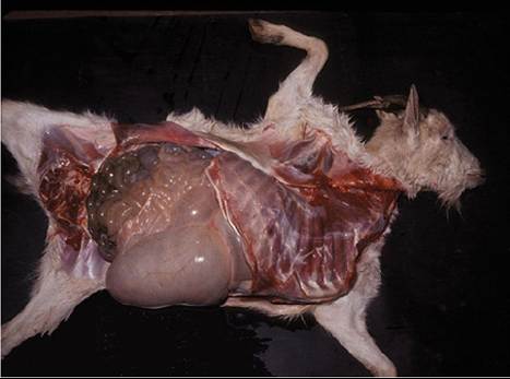
Figure 16.1 Emaciated goat lacking omental fat. The intestines should not be easily seen until the omentum has been removed.
Source: Courtesy of Dr. M.C. Smith.the rib cage. Foot trimmers permit cutting of the ribs at the costochondral junction, even in adults. The rib cage of a kid is folded back as one section. In an old goat, it may be necessary to use heavy tree-branch loppers or to cut between the ribs and break one at a time near the spinal column. Examine the viscera in the thorax and abdomen systematically and note any fluid accumulations.
The brain of a young kid can be exposed by cutting the calvarium with heavy scissors or foot trimmers, but a saw or hatchet is needed when removing the brain of an adult. Remove skin from the top of the skull and make a first transverse saw cut just caudal to the zygomatic process of the frontal bone. Then make sagittal cuts on each side from just medial to the occipital condyle to join the first cut (King et al. 2005). Alternatively, four saw cuts can be made in a diamond pattern, with the anterior point of the diamond on the midline between the eyes. If the goat has large horns, the first transverse cut is made on the forehead anterior to the horns and angled caudally. The sagittal cuts are slanted toward the midline, and the skull cap with horns can then be lifted off, as illustrated in Figure 16.2. Don't forget to slit the cheeks and examine the molar teeth before discarding the skull.
Heart blood, urine (50 mL if possible), aqueous humor, and cerebrospinal fluid (at the atlanto-occipital space) can be aspirated in a clean fashion and set aside for later examination. For instance, glucose is usually present in the urine of a goat that dies of enterotoxemia and might be found in aqueous humor (Debien et al. 2013). Unfortunately, information regarding sensitivity and specificity of various findings in specified reference populations - for example, the percentage of goats dying in New York State with enterotoxemia that have glucosuria (equals sensitivity) or the
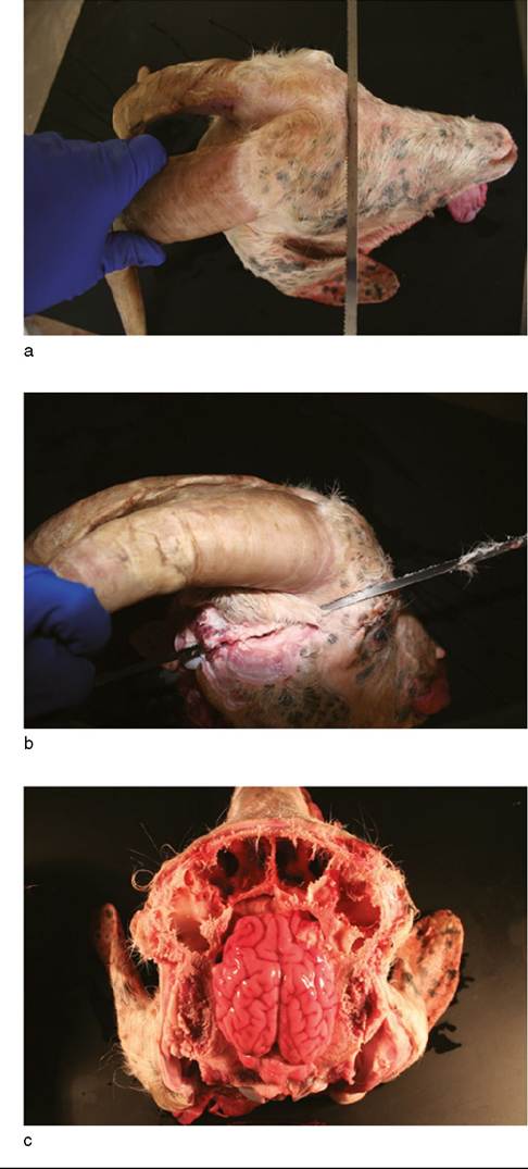
Figure 16.2 Opening the cranial cavity of a horned goat.
(a) The initial transverse saw cut is made on the forehead in front of the horns. (b) The sagittal cuts are almost horizontal.
(c) The horns and top of the calvarium have been lifted off to expose the brain and frontal sinus. Source: Courtesy of Dr. M.C. Smith; Mr. Patrick Burke, prosector.
percentage dying of something other than enterotoxemia that do not have glucosuria (equals specificity) - is rarely available. Until the published literature provides such
information, practitioners should tabulate their own records as to gross necropsy findings, simple test results, and final diagnosis. This permits later estimation of sensitivity, specificity, and even prior probability in a given practice setting.
Appropriate tissues or swabs (e.g., abnormal lung, mammary gland, abscesses, or tied-off section of intestine) should be taken for bacteriologic and virologic testing. The laboratory to receive the specimens should be consulted relative to transport media, packaging, and shipping instructions. Sections should be taken for histologic study from the major organs (i.e., lung, liver, kidney, heart, mesenteric lymph node, brain stem, cerebellum, and cerebrum) and from all suspected lesions. The kidney section should include cortex, medulla, and pelvic epithelium. Whenever possible, some normal tissue at the edge of a lesion should be included. Slices must be thin (no more than 5 mm) and the tissues should be preserved in at least 10 times their volume of 10% formalin. A history should accompany the samples to the laboratory. Saving tissues preserved in formalin is advisable, even if the owner does not currently wish to pay for histologic examination.
The gastrointestinal tract should not be opened until the rest of the necropsy is finished, to avoid contamination. At the appropriate time, rumen contents should be examined for pH level, color, smell, and identifiable roughages or grain. A sample of at least 200 g should be labeled and frozen for later reference if toxicosis has not yet been ruled out. At the same time, large pieces (at least 100 g each) of liver and kidney should be frozen (King 1983). Liver also may be useful for monitoring the trace mineral status of the herd. Plant fragments requiring identification should be dried between layers of newspaper. The abomasum and small intestine should be examined closely for parasites; this serves a herd-monitoring function, even if parasitism did not directly kill the goat (Figure 16.3). A quantitative
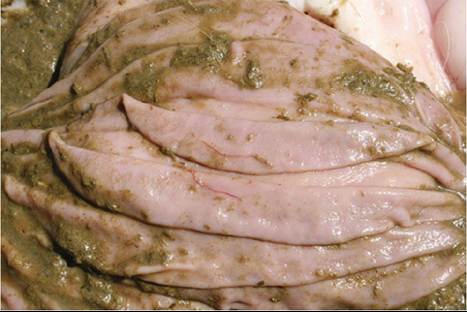
Figure 16.3 Two Haemonchus contortus worms with barberpole appearance are present on the abomasal folds. Source: Courtesy of Dr. M.C. Smith.
fecal examination may be warranted. Peyer's patches and mesenteric, bronchial, and mediastinal lymph nodes deserve special attention, and the presence or absence of white foci in the wall of the intestine (coccidiosis) should be determined. Impression smears of intestinal mucosa may be helpful for diagnosis of coccidiosis, cryptosporidiosis, paratuberculosis, and (if the necropsy is done less than four hours after death) enterotoxemia.