Swellings Involving the Abdomen and Escutcheon
Alteration in the abdominal silhouette is discussed in detail in Chapter 10.
Umbilical Hernia
Umbilical hernia caused by the incomplete closure of the umbilical ring (in the absence of omphalitis) is rare in goats.
A genetic predisposition has not been identified in this species (Hamori 1983). Nevertheless, prudence dictates that males that will be used extensively for breeding should be free of all recognizable congenital defects.Goats with a swelling originating from the umbilical region should be examined by palpation while standing, and if necessary in lateral recumbency. Ultrasound examination will reveal abomasum or loops of intestine, if present. If the swelling is easily reduced with no tenderness or thickened stalk, then it is likely that it is a her - nia unaccompanied by an abscess. In young female goats, wrapping the abdomen for two to four weeks with adhe - sive elastic tape may permit closure of the hernia ring to occur. A commercial cloth back brace, women's size, might be used instead of the tape and would be more eas - ily adjusted as the animal grew. If the defect is large, an internal abscess is suspected, or the patient is male, surgical repair is the preferred therapy. The goat is held off feed and placed in dorsal recumbency and standard techniques appropriate for herniorrhaphy in calves are employed (Ortved 2017). Prosthetic mesh may be required to close large defects (over 3 cm) in the body wall (Fubini and Campbell 1983). Successful closure of umbilical hernia rings 7-10 cm in diameter has been achieved in goats using double-layer polypropylene mesh (Vilar et al. 2011).
An alternative non-surgical repair has been described using an elastrator castration band. The female animal is lightly sedated and placed in dorsal recumιbency With all of the contents of the hernia, sac replaced into the abdomen, two metal pins such as diaper pins are inserted through the skin on each side of the sac, so that they will sit close to the abdominal wall.
An elas - trator band is then placed around the hernial sac, between the pins and the body wall, where it causes sloughing of the skin and healing of the defect within two weeks (Navarre and Pugh 2002). Tetanus prophylaxis is imperative.UmbiLicalAbscess
If palpation of an umbilical swelling reveals warmth, tenderness, or just an irreducible fluctuance, diagnostic aspiration is indicated. Omphalitis may result in abscessation of remnants of umbilical arteries, the umbilical vein, or the urachus. Ultrasonography may be useful for evaluation of the extent of internal involvement of an umbilical abscess. External abscesses are drained and systemic antibiotics are administered for one week or longer. If evidence of infection or concurrent hernia persists, surgical debridement and herniorrhaphy are recommended.
Abdominal or Flank Hernia
The abdominal wall of a goat is relatively thin. Muscle tearing and separation often occur from blunt trauma during fighting, shearing, or crowding through narrow doorways. Diagnosis of a unilateral flank hernia is usually obvious, although it is important to determine if a preg - nant uterus is trapped in a subcutaneous location (hys- terocele). The hernia ring may be located some distance dorsal to the subcutaneous swelling (Abdin-Bey and Ramadan 2001). When the hernia or stretching of the body wall is bilateral (as from trauma from doorways) it should be distinguished from pregnancy, hydrometra, obesity, or ascites by careful physical and ultrasound examination.
Small hernias may be surgically repaired, while hernias large enough to impede parturition are usually an indication for culling in a commercial herd. Tranquilization with xylazine and local anesthesia using a ring block of lidocaine (not to exceed 10 mg/kg) has been used for repair of abdominal hernias in goats (Al-Sobayil and Ahmed 2007). Either absorbable or non-absorbable suture is satisfactory, with non-absorbable (the authors used silk) favored in older animals or those with more longstanding hernias.
Ventral Hernia
Occasionally, trauma or extreme abdominal distention leads to rupture of the ventral abdominal muscles caudal to the umbilicus, as described in sheep (Arthur et al. 1989). This results in edematous swelling of the abdominal wall and dropping of the udder. Successful surgical repair has been reported in a goat (Misk et al. 1986). The late pregnant uterus can become trapped in the hernia in a subcutaneous location, making vaginal delivery difficult (Horenstein and Elias 1987). Successful surgical correction of such a “metrocele” or hysterocele has been reported several times, including by Radhakrishnan et al. (1993) and Vijayanand et al. (2009).
Inguinal and scrotal hernias are discussed in Chapter 13.
Ventral Edema of Angoras
Angora goats in South Africa, the United States, New Zealand, and England have developed severe edematous swelling (“swelling disease” or “water belly”) of the ventral abdomen and chest and sometimes the limbs and submandibular region after shearing or other stress (Mitchell et al. 1983; Byrne 1994a, b; Thompson 1994). As many as 15% of the herd may be affected. The swelling consists of clear, copious, non-clotting fluid and disappears spontaneously in a few days. Possible etiologies discussed in the literature include hypoproteinemia and change in capillary permeability, stress (via increased aldosterone secretion and sodium retention), and vitamin E deficiency. Research attempting to recreate the condition with parasitic infections or modify it with increased dietary protein has been inconclusive, but in general the smaller goats with lower plasma total protein concentrations are at higher risk (Snyman and Snyman 2005). As the condition is most common in highly bred Angoras 4-18 months old, inbreeding should be avoided, in addition to correcting factors that predispose to hypoproteinemia (Bath et al. 2005).
Hypoalbuminemia due to parasitism or paratuberculosis and edema caused by congestive heart failure must be ruled out in individual animals that do not recover rapidly.
If sudden deaths occur in the herd, vitamin E status should be investigated (Byrne 1994a). Otherwise, no treatment is indicated except provision of feed and shelter for newly shorn animals.Urethral Rupture
Bucks and wethers with urolithiasis may develop a ventral swelling if the urethra ruptures and urine leaks into subcutaneous tissues. The swelling is edematous and cool; aspiration yields a watery fluid that may have an ammonia- cal odor when heated and its urea nitrogen concentration will be high. The fluid can be reabsorbed if the obstruction to normal urine flow is relieved. Localized skin sloughing may occur. Urolithiasis and its treatment are discussed in detail in Chapter 12.
Gangrenous Mastitis
The skin of the ventral abdomen anterior to the udder may become swollen and edematous because of vascular thrombosis in goats with gangrenous mastitis. Initially the swelling is cool; necrosis and sloughing may eventually occur if the animal survives. Gangrenous mastitis is discussed in Chapter 14.
Ectopic Mammary Gland
The embryonic mammary line of mammals extends from the pectoral region to the vulva. Although female goats usually develop only two functional glands in an inguinal location, milk-secreting tissue is occasionally located bilaterally in the lips of the vulva (Lesbouyries and Drieux 1945; Kulkarni and Marudwar 1972; Ramadan and El Hassan 1975; Smith 1986). As parturition approaches, the vulva enlarges, as does the udder. This vulvar swelling is firm and lobular (Figure 3.12), is separated from the skin, and does not subside promptly after parturition as physiologic edema would. Instead, the vulva remains distended for as long as three months, but eventually subsides as the glandular tissue becomes atrophied from the back-pressure of entrapped milk. The condition is merely a curiosity, but can be confirmed by aspiration of a whitish fluid containing fat globules. The bilaterally symmetrical nature of the swelling also helps to differentiate ectopic mammary gland from an adenocarcinoma or other neoplasm (see Chapter 2).
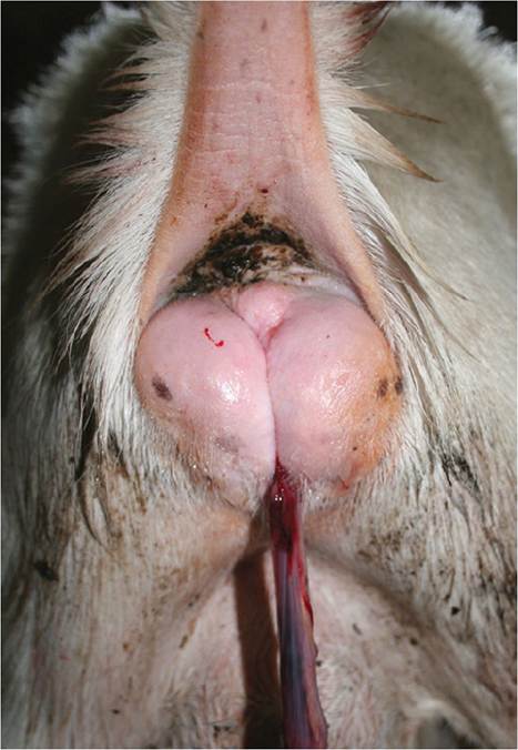
Figure 3.12 Ectopic mammary gland distending the vulva of a mature Saanen doe one day after parturition. The doe also has a retained placenta. Source: Courtesy of Dr. M.C. Smith.
Perineal hernias have been reported occasionally in goats (Abdin-Bey and Ramadan 2001) and can produce a swelling in the ischiorectal fossa lateral to the vulva and anus, but would be unilateral.
Mammary gland development in the buck (gynecomastia) is discussed in Chapter 13.