Swellings Involving the Neck and Chest
Caseous lymphadenitis, as already discussed, should always be considered when swellings are present in the region of cervical or prescapular lymph nodes. Many other conditions of varied etiology cause diffuse or localized swellings on the neck or chest.
Abscesses OtherThan Caseous Lymphadenitis
Abscesses that do not respond to simple drainage should be reassessed (including radiographic examination) for the possible presence of a foreign body such as a needle, plant fragment, or piece of necrotic bone. Abscesses in the neck and near the left elbow have been reported in goats that ingested sewing needles (Sharma and Ranka 1978; Tanwar and Saxena 1984).
Mycetomas, as discussed in Chapter 2, are chronic, subcutaneous, fungal-induced swellings, often with underlying periosteal reaction.
Tissue Necrosis (“Sterile Abscesses”) Caused by Injections
A number of vaccines, including multivalent clostridial (Green et al. 1987), paratuberculosis (Holstad 1986d), caseous lymphadenitis, and foot and mouth disease vaccines, cause large, roughly spherical, firm, persistent swellings (granulomas). This occurs in most of the animals vaccinated and does not require contaminated vaccine or dirty needles to occur. A provisional diagnosis is based on the presence of such a tissue reaction in a site known to have been used previously for vaccination. No treatment is required in commercial animals unless local infection occurs. These swellings are of more concern in pets and show goats, where surgical extirpation of the lump may be requested by the owner. It is important to inject these animals in a cosmetically acceptable location, such as behind the shoulder or elbow, because a reaction can be expected from many adjuvanted, effective vaccines. In general, injections should not be given subcutaneously in the neck of a show animal. It is difficult for veterinarians writing health certificates to feel confident that goats with a nodular swelling in this region are free of caseous lymphadenitis.
Administering vaccines and antibiotics deep into musculature is not a satisfactory method of avoiding problems. The result, an often severe necrosis of muscle, is more difficult to observe, but clearly painful to the goat. Tender, firm swellings, lameness, or reluctance to rise are common. When a site in the hindlimb is used, sciatic nerve paralysis is also common, especially in young or emaciated animals with limited muscle volume. In addition, if the animal is to be used for meat, the muscle necrosis, which outlasts tissue residues by weeks or months, results in carcass damage that requires trimming or that escapes the notice of the meat inspector, but is obvious and unappealing to the consumer.
Certain solutions are so irritating to tissue that they should not be administered subcutaneously or intramuscularly to goats. Some proprietary calcium preparations with phosphorus and dextrose included are noted for causing sloughing of tissue at the injection site. Failed attempts at intravenous injection may cause large swellings over the jugular vein where irritating drugs were mistakenly deposited in a perivascular location.
In animals kept for antibody production, it is normal to find numerous firm subcutaneous nodules where the adju- vanted antigen has been injected.
Wattle Cyst and Thyroglossal Duct Cyst
Wattles are reportedly caused by a dominant autosomal gene with complete penetrance but variable expression regarding the shape and location of the wattles (Ricordeau 1981). Occasionally, cysts occur unilaterally or bilaterally at the base of the wattle or at the site of previous wattle amputation. These cysts are certainly not rare, but rarely are of enough concern to be reported in the literature. They are variously referred to as branchial cleft cysts (Williams 1980), dermoid cysts (Gamlem and Crawford 1977), wattle cysts (Fubini and Campbell 1983), or tassel cysts. Wattle cysts are usually present at birth, although they may enlarge with time and become more noticeable later.
They contain a thick or thin, clear liquid and refill (Abu-Seida 2014) or become abscessed after aspiration. They can be excised intact under local anes - thesia if care is taken to avoid the underlying jugular vein; excision may be necessary in show goats because of possible confusion with caseous lymphadenitis. Breed predilections reported in the literature may be biased by the prevalence of given breeds or family lines in certain regions.Wattle cysts should be differentiated from thyroglossal duct cysts that are located on the midline, below the hyoid apparatus (Al-Ani and Vestweber 1986; Nair and Bandopadhyay 1990; Al-Ani et al. 1998). Thyroglossal cysts sometimes become quite large over time and can be removed surgically with due care.
Thyroid Gland and Goiter
Anatomy and Physiology
The thyroid of the goat is bilobed and located slightly behind the larynx. Right and left lobes, which lie lateral to the trachea, are joined by a thin isthmus that passes across the ventral aspect of the trachea (Reineke and Turner 1941). In young animals, the lobes of the thyroid gland are often embedded in thymic tissue. The isthmus becomes more fibrous (less glandular) and more caudally located with advancing age (Roy et al. 1975).
The thyroid gland forms several hormones by iodinating organic compounds that contain the amino acid tyrosine. Thyroxine (T4) and 3,5,3-triiodothyronine (T3), when formed, are stored in colloid within the acini of the thyroid gland until needed. The thyroid gland and its hormones control the metabolic rate of an animal by regulating cellular oxidation (Wilson 1975). Selenium is required for hepatic conversion of T4 to T3 (Kohrle 2000).
Goiter is an enlargement of the thyroid gland. In ruminants, goiter usually suggests attempted compensation for a hypothyroid state. Normally, low thyroxine and triiodothyronine output stimulates increased thyrotropin (thyroid-stimulating hormone) output, which leads to increased iodine uptake from the blood and hyperplasia of the gland.
The enlarged gland may be able to compensate by increased iodine trapping.Normal Thyroid Function Tests
Natural hypothyroid states other than goiters have not been reported in goats. If testing is done, it should be remembered that, at least in dogs, corticosteroid therapy and the terminal stages of non-thyroidal illnesses can both depress plasma T4, possibly because of interference with T4 binding by plasma proteins.
The thyroid function of goats has been investigated by researchers wishing to use the species as a model for the physiology of larger ruminants. For instance, uptake of radioiodine (131I) has been studied in goats (Flamboe and Reineke 1959; Davis et al. 1966; Ragan et al. 1966). Thyroidectomy, as discussed below, has also been performed to study normal thyroid function.
Thyroid hormone level determinations are more accessible to practitioners, although caprine normals are not well established. Repeated serum thyroxine level determinations in 20 female and 40 male goats of dairy breeds, from 2 weeks to 6 years of age, yielded a mean of 6.53 ± 0.03 (standard error, SE) μg∕dL of thyroxine. The range was 2-17 μg∕dL (Anderson and Harness 1975). Thyroid function tests were also performed on up to 55 pygmy goats from a laboratory colony (Castro et al. 1975). No sex differences were noted. Mean values recorded included proteinbound (organic) iodine, 8.1 ± 1.2 (standard deviation, SD) μg∕dL; T4, 7.2 ± 1.1 μg∕dL; and cholesterol, 90 ± 29.7 mg∕ dL. Because values were within normal human ranges, the authors concluded that there was no evidence of thyroid malfunction.
As part of a study to establish normals for 10 species, thyroid hormones were assayed in duplicate from 10 goats (Reap et al. 1978). Average values (± SD) and ranges were T4, 3.45 ± 0.47 (3-4.23) μg∕dL and T3, 145.9 ± 29.32 (88-190) ng∕dL. The average thyroxine value for four young and four adult goats (breed not specified) was 4.25 μg∕dL in another study (Kallfelz and Erali 1973).
Various nutritional, seasonal, and endogenous influences on thyroid hormone levels in goats and sheep have been reviewed by Todini (2007). A few goat studies are described here. Thyroxine levels in Angoras in South Africa were found to fluctuate with no obvious seasonal pattern (Wentzel et al. 1979). In another study of 70 female and 10 male indigenous goats in Italy, T3 and T4 levels were highest in the spring and lowest at the end of the summer; the mean decrease in T3 was 57% and in T4 it was 35%. Older goats also had lower levels than did goats under 3 years of age (Colavita et al. 1983). For Angora goats in Turkey, T3 and T4 levels were found to be lower under warm environmental conditions and higher during cold months (Pehlivan and Dellal 2017). In one study of dairy goats, the plasma thyroxine level decreased by approximately 30% during the first week of lactation, relative to the prepartum period (Emre and Garmo 1985).
The author has evaluated thyroid-stimulating hormone response in three normal adult dairy goats (M.C. Smith, unpublished data). Serum thyroxine concentrations approximately doubled four hours after intravenous injection of 5 IU of thyrotropin. A similar response was seen in 25 juvenile goats after receiving intravenous thyrotropinreleasing factor at 1 μg∕kg; T3 increased a mean of 318% at one hour and T4 increased a mean of 174% (97-227%) at four hours after stimulation (Reinemeyer et al. 1991). Baseline values in this study were 30-90 ng∕dL T3 and 3.1-6.1 μg∕dl T4.
Experimental Hypothyroidism
Thyroidectomy has been performed in the goat. Ectopic thyroid tissue can be destroyed by 131I administration where surgical removal of the visible thyroid glands is not sufficient (Ekman 1965). Parathyroid function is maintained because a pair of external parathyroids is located separately from the thyroid glands, embedded in thymic tissue.
When adult goats underwent thyroidectomy, the major clinical sign was bodyweight gain. Does that received 131I during pregnancy produced stillborn kids with no detectable thyroid glands. These does had reduced milk production. Goats that underwent thyroidectomy developed marked thickening and softening of the skin with local hair loss and they shivered intensely when exposed to cold (Andersson et al. 1967).
When young kids underwent thyroidectomy, growth stopped within one to two months, kids became lethargic, and the head developed a dish-faced appearance (Reineke and Turner 1941). Growth recommenced if iodinated proteins were fed.
Goats with hypothyroidism have also been created experimentally by daily oral feeding of thiourea (Sreekumaran and Rajan 1978) and by injection of methyl thiouracil or thiourea (Gupta et al. 1990). When young kids were thus treated, weight loss and myxedema (edema and gelatinous infiltration of subcutaneous tissue) developed (Sreekumaran and Rajan 1977). Skin changes in these kids are discussed in Chapter 2. When 8-month-old male goats were made hypothyroid with thiourea, plasma testosterone levels decreased to less than 12% of pretreatment levels (Gupta et al. 1991). Long-term feeding of Brachiaria mutica grass with high but sublethal nitrate concentration has resulted in abnormally small and firm thyroid glands and mild proliferation of the acinar epithelium (Prasad 1983).
Naturally Occurring Dietary Goiter
Enlargement of the thyroid gland is usually a nutritional disease.
Etiology There are two major mechanisms by which diet can cause development of goiters in goats: iodine deficiency and feeding of goitrogens. In addition, experimental cobalt deficiency (and thus vitamin B12 deficiency) has produced elevated thyroxine levels accompanied by marked hypertrophy and hyperplasia of the thyroid gland (Mgongo et al. 1981); however, in a later study thyroid function was not markedly impaired by cobalt deficiency (Mburu et al. 1994).
Iodine-Deficiency Goiter “Endemic” goiter in goats has generally been recognized in the past in the same regions where human goiter occurred. Soils are deficient in iodine or do not release it readily to plants and drinking water. Iodine-deficient regions include the Great Lakes, Great Plains, Rocky Mountains, and Pacific Coast regions of the United States (Lall 1952), Switzerland, parts of Great Britain (Wilson 1975), and the Himalayas (Raina and Pachauri 1984). The availability of iodized salt has decreased the prevalence of this type of goiter in both the United States and Western Europe. Goiter remains common in the Himalayas, where supplementation is not as available (Singh et al. 2002).
Breeds of goats vary in sensitivity to iodine deficiency. The Boer goat, a rapidly growing meat breed from South Africa, seems to be especially susceptible. It is possible that selection for resistance to iodine deficiency would be selection for slow growth rate (van Jaarsveld et al. 1971). In the Himalayas, where caprine goiter was extensively studied, indigenous strains were apparently more resistant to iodine deficiency than were goats (Barbari or Alpine) purchased from outside regions (Rajkumar 1970). The Angora goat is apparently very susceptible to iodine deficiency (Kalkus 1920).
Goitrogens Goitrogens are compounds that interfere with the uptake of dietary iodine or with its metabolism in the formation of thyroxine (Bath et al. 1979; Cheeke 1998). Thiocyanate is a goitrogen present in many species of brassicas, members of the Brassicaceae (Cruciferae) family. Increasing dietary iodine will overcome the inhibitory effects of thiocyanate on selective concentration of iodine by the thyroid. Goitrin (thiooxazolidone) is a thiouracil- type goitrogen found in the seeds of rape, kale, and other Brassica spp. that blocks hormone synthesis; its action cannot be overcome by feeding additional iodine.
Thiourea is similar and also interferes with organic binding of iodine; it has been used to produce experimental hypothyroidism (Wilson 1975). Mimosine in L. Ieucoceph- ala is broken down metabolically to a goitrogen in ruminants (Hegarty et al. 1976; Prasad 1989), but this chemical can be detoxified by natural populations of rumen bacteria (Hammond 1995). Long-term consumption of Leucaena has caused alopecia and colloid goiter in a goat in Brazil (Peixoto et al. 2008) and adult and fetal goiters in India (Sastry and Singh 2008; Reddy et al. 2016). See further discussion in Chapter 10. African pearl millet (Pennisetum typhoides) is goitrogenic for goats, although the toxin has not been identified (Abdel Gadir and Adam 1999).
Clinical Signs and Laboratory Findings The normal thyroid gland is 0.20% of bodyweight (Kaneko 1997). In goitrous regions, unsupplemented adults have marked enlargement of the thyroid, which is sometimes the size of an orange (Kalkus 1920). The goats otherwise look healthy. Reproductive performance may be decreased (Kategile et al. 1978). Humoral and cell-mediated immune responses may be decreased (Singh et al. 2006).
Affected kids have thyroid enlargement at birth (Figures 3.8 and 3.9), and enlargement of the kid's pituitary gland has also been described (Ozmen and Haligur 2005). They may be stillborn or are very weak and die within a few hours. Sometimes the goiters are large enough to cause dystocia (Reddy et al. 2016; Singh et al. 2019). Increased blood flow to the thyroid may cause a palpable thrill. Most of these kids are hairless or covered with very fine hair, while some have a normal hair coat (Kalkus 1920; Love 1942; Paliwal and Sharma 1979). It has suggested that multiple fetuses in a litter are more apt to be affected with dietary goiter than is a single fetus (Ozmen and Haligur 2005).
In one outbreak in South African Angoras, believed to be caused by thiocyanate from heavily fertilized alfalfa pastures, kids were viable but abnormal from birth (Bath et al. 1979). They were short, blocky, obese, and inactive. The skull was broadened laterally and prognathia inferior was present in all cases. Goiters were easily palpated bilaterally. Mean thyroxine level for four goitrous goats was 3.1 gg/dL, whereas the mean of four normal goats was
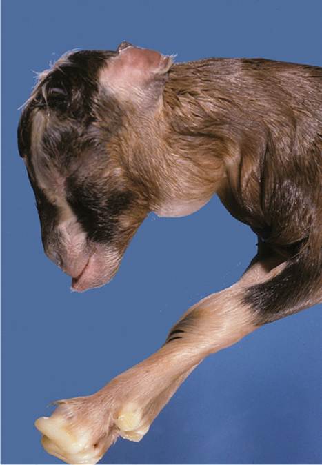
Figure 3.8 Congenital iodine-deficiency goiter in a stillborn kid. Source: Courtesy of Dr. M.C. Smith.
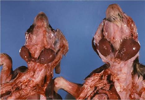
Figure 3.9 Goiters exposed in twin stillborn kids by reflecting the skin. Source: Courtesy of Dr. M.C. Smith.
5.9 gg/dL. Plasma cholesterol was five times higher (10.9 mmol/L versus 1.8 mmol/L) in goitrous than in control goats. In another case report, does were fed on cabbages and the kids appeared normal, except for goiters measuring as large as 7 cm ? 4.5 cm (Lombard and Raby 1965). The largest goiters were associated with severe dyspnea and death, whereas less markedly affected kids grew poorly.
A study from an iodine-deficient area of India evaluated 252 congenitally goitrous kids born to 574 does with goiter. Clinical signs included thyroid gland hypertrophy and palpable thrill, enlarged joints, muscle contracture, an arched back and waddling gait, partial to complete alopecia, weakness, and lethargy. Hydrocephalus and prognathism were less common. Serum cholesterol was increased while T3 and T4 were decreased. The incidence of stillbirth in this study was 18% (Singh et al. 2003).
Diagnosis Major differentials for a diagnosis of goiter include encapsulated abscesses and wattle cysts, which are unlikely to be bilateral and in the exact location of the thyroid. Thymic enlargement is more difficult to distinguish because the thyroid glands are normally embedded in the thymus. The kid with an enlarged thymus grows rapidly, has a healthy haircoat, and is active. As long as trace mineralized salt is available and no known goitrogens are being fed, the continued good health of the growing kid remains the ultimate diagnostic test to rule out goiter. Biopsy is not necessary, although normal thyroids contain acini lined by low cuboidal epithelium and filled with colloid, as opposed to the tall columnar epithelium, papillary infolding, and minimal colloid of goitrous glands (Love 1942; Roy et al. 1964). A hyperplastic goiter is transformed into a colloid goiter when the diet is improved or the lower iodine needs of an older animal are met, resulting in a gland that is still enlarged but has much colloid in the acini. Iodine content of the goitrous thyroid gland is reduced. Subclinical goiter is diagnosed when the glands appear to be normal size but are histologically hyperplastic.
Treatment and Prevention The actual dietary iodine requirement for goats, as discussed in Chapter 19, is 0.8 mg/ kg dry matter for lactating females and 0.5 mg/kg for the rest of the herd. Cruciferous plants increase the ration iodine requirement to approximately 2 mg/kg.
Iodine-deficiency goiter is treated or prevented by supplying iodine to the goat, especially the pregnant doe. This can easily be done with iodized salt, assuming that no iodine-free salt source is available for the goats to satisfy their salt requirements. In a report from India, biochemical and hormonal values normalized in goitrous kids when colloidal iodine (I2) was given orally at 0.1 mg/kg bodyweight for 100 days (Singh et al. 2003). Synthetic sodium thyroxine (0.2 mg/day orally to goats weighing 16-20 kg) also corrected many clinical signs of deficiency (Singh et al. 2006), but should not be necessary once the diet is corrected.
In the classic experiments of Kalkus (1920), oral daily potassium iodide (2 grains, 130 mg) or weekly application of 1 mL of tincture of iodine to the back throughout gestation were both successful in preventing goiter. Iodine treatment during the preceding pregnancy sometimes was enough to permit production of another normal kid. Adult goats with iodine-deficiency goiter showed a decrease in thyroid size after treatment (Kalkus 1920; Welch 1928). Goat kids with goiters should benefit from topical application of iodine, as with navel dips. In some countries, injectable iodized poppy seed oil is available and provides supplementation lasting for up to two years (Wichtel et al. 1996).
Congenital goiter caused by goitrogens is best avoided by not feeding the incriminated forages (especially brassicas) during pregnancy. Supplemental iodine can also be fed to the does (de las Heras et al. 1984).
Hereditary Goiter
Congenital goiter occurred spontaneously in an inbred strain of Dutch goats (mixed Saanen and dwarf goats). The trait was maintained and studied extensively as a model for thyroid defects in humans.
Pathogenesis The condition was inherited as an autosomal recessive trait (Kok et al. 1987). Thyroglobulin, the normal precursor of the thyroid hormones T3 and T4, was not produced in the goat that was homozygous for this trait. As a consequence, the normal feedback mechanisms were impaired and continuous thyrotropin secretion led to development of a goiter. The responsible mutation in the thyroglobulin gene has been characterized (Rivolta and Targovnik 2006).
Clinical and Laboratory Findings Normal thyroids (both together) in this breed weighed 1-4 g, and goiters weighed 15-300 g. Plasma T3 levels (9-36 ng/dL) and T4 levels (less than 0.4 pg/dL) were substantially lower than normal goat T3 (124-151 ng/dL) and T4 (5.9-10.2 pg/dL) levels (de Vijlder et al. 1978). Histologically, the goitrous thyroid had hypertrophic and hyperplastic epithelium consistent with prolonged thyrotropin stimulation. Colloid was almost absent.
In addition to having enlargement of the thyroids, goitrous kids were sluggish and grew poorly. The haircoat was rough and sparse. If thyroxine replacement was not provided, the haircoat eventually disappeared almost completely and the skin became thick and scaly (Rijnberk 1977). Euthyroidism could be achieved with these goats using iodide supplementation (1 mg I- per day orally). Other proteins were iodinated and then converted to T3 and T4, even though thyroglobulin still was not synthesized (van Voorthuizen et al. 1978).
Congenital Goiters in Other Breeds Congenital goiters of possibly hereditary etiology have also been found in Boer goats (van Jaarsveld et al. 1971). Except for the enlargement of the thyroid glands (average 37 g compared with the normal average 2 g), the kids appeared normal. Histologically, there was hypertrophy of the thyroid epithelium with absence of colloid. Normal iodoproteins were present, but the thyroglobulin polymers tended to dissociate into subunits. Congenital goiters (average 43 g compared with a normal average of 9 g) have also been suspected to be hereditary in Shami dairy goats (Al-Ani et al. 1998). An autosomal recessive hereditary goiter in goats in Inner Mongolia was controlled by removing known carriers and identifying other carriers in the herd using a goat-homologous assay for thyrotropin (Mei and Chang 1996).
Thymus
Young kids, especially if well fed, sometimes develop bilaterally symmetric swellings in the upper neck in the region of the thyroid gland (Figure 3.10). The swellings may first become noticeable as early as 2 weeks of age and regress spontaneously, often at about 4 months of age (Pritchard 1987) or somewhat later (Bertone and Smith 1985). The kid is otherwise healthy, with a good haircoat, and therefore is unlikely to be affected with severe iodine deficiency or goiter. Another subcutaneous swelling often occurs at the thoracic inlet. Aspiration of either the
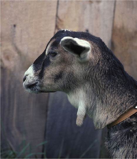
Figure 3.10 Enlarged thymus in the upper neck of a rapidly growing Nubian cross kid. Source: Courtesy of Dr. M.C. Smith.
cranial or caudal cervical masses, while not indicated, yields thymic tissue. The origin of the excessive thymic tissue might be remnants from embryologic development or accessory thymus; in any case, the glandular tissue involutes naturally and no treatment is needed. Owners should be warned of the potential for iodine toxicity if unnecessary supplements, such as kelp, are fed when no thyroid deficiency exists.
Tumors of the thymus occur in adult goats, and their presence as a space-occupying lesion or incidental finding in the thorax is discussed in Chapter 9. Less commonly, remnants of the cervical portion of the thymus become neoplastic. Diagnosis is by examination of a biopsy or aspirate, and treatment by surgical excision after ultrasound examination for surgical planning and radiographic evaluation for possible presence of tumor masses within the thorax (Hill et al. 2017).
Thymic tissue may also be embedded within the thyroid gland of goats of any age (Roy et al. 1976), often in association with parathyroid tissue, but no clinical relevance has been reported for this histologic finding.
Phlebitis
Because the jugular veins are commonly used for venipuncture and administration of various medications,
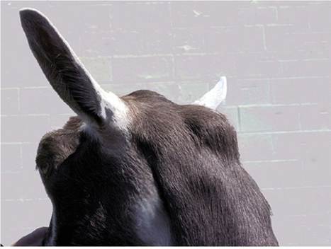
Figure 3.11 Distension of the atlantal bursa in a goat with clinical caprine arthritis encephalitis. Source: Courtesy of Dr. M.C. Smith.
iatrogenic phlebitis may be expected to occur occasionally. Hematomas typically resolve quickly without treatment, whereas perivascular deposition of irritating drugs can lead to cellulitis or even abscessation. Conservative therapy, including application of hot compresses, is preferable to lancing a swelling so close to the jugular vein.
Atlantal Bursitis
Atlantal bursitis is a fairly specific but uncommon sign of caprine arthritis encephalitis virus infection. A fluctuant swelling is located underneath and extends on both sides of the ligamentum nuchae (Figure 3.11). The bursa often contains mineralized material, which can be demonstrated with a radiograph (Garry and Rings 1985). Histology shows hyperplasia of synovial cells and mononuclear cell infiltration (Gonzalez et al. 1987). An abscess can develop if aspiration is attempted. The supraspinous bursa may be similarly involved, as may be other bursae over the carpus, olecranon, or tuber ischii. Usually at least some degree of lameness is present by the time the atlantal bursa becomes noticeably distended. See Chapter 4 for further discussion of the virus.
SternalAbscesses and Hygromas
Sternal abscesses were detected in 72 goats in a large antibody-producing herd over a 16-year period (Gezon et al. 1991). These abscesses were 3-15 cm in diameter and usually involved skin and subcutaneous tissues, but rarely invaded muscle or bone. Two of the goats, however, had osteomyelitis of the sternum. Bruises and abrasions of an area commonly exposed to manure were proposed as an explanation of the occurrence of the abscesses. Antibiotic therapy had little effect on the condition. The authors did not adequately report culture results, and thus no conclusions can be drawn from this study concerning bacteria present in unbroken sternal abscesses.
Sternal abscesses are, in the authors' experience, most common in goats afflicted with severe lameness, such as from arthritis associated with caprine arthritis encephalitis virus infection. Two hypotheses come to mind to explain this association. First, the lame goat spends more time in sternal recumbency and therefore may develop a hygroma or decubital sore. Second, the caprine arthritis encephalitis virus-induced arthritis might involve joints between stern- ebrae, with secondary bacterial infection. Successful treatment of these abscesses often requires surgical debridement. Radiographic or computed tomography evaluation for osteomyelitis or enlargement (and probable infection) of lymph nodes within the thoracic cavity is helpful in offering a prognosis and determining the extent of surgical debridement required. Goats that are markedly lame and emaciated or have extension of infection into the thorax will probably benefit little from treatment of a sternal abscess.
Warbles
Larvae of Przhevalskiana silenus, a warble fly that migrates to the back region of goats in Mediterranean countries and Asia, are accompanied by localized inflammatory reaction and granulation tissue in the subcutaneous tissue. This parasite is discussed in Chapter 2. Although cattle warble fly larvae (Hypoderma spp.) could potentially invade goats, natural occurrence has not been documented and experimental infections with Hypoderma Iineatum larvae did not progress to the development of subcutaneous warbles (Colwell and Otranto 2006).
Tapeworm Cysts
The intermediate form of the dog tapeworm Taenia multi- ceps (previously called Multiceps multiceps or M. gaigeri) forms cysts within the central nervous system (CNS) of sheep and goats, but also outside the CNS of goats, especially in muscle and subcutaneously (Verster 1969; Schuster et al. 2010). Scolices are arranged in clusters and there are no internal or external daughter cysts. These cysts have been reported as the cause of large (approximately 15 cm in diameter) subcutaneous swellings distributed over the limbs and body of goats in the Sudan (Ramadan et al. 1973; Hago and Abu-Samra 1980) and India (Shastri et al. 1985). The cysts are fluctuating, cool, and covered with hairless skin. Depending on the cyst location, there may be interference with locomotion, feeding, or function of internal organs. Head shaking is commonly observed when cysts are near the base of the ear.
Several methods of treatment have been successful, including excision of the cyst, lancing the cyst and packing the cavity with gauze soaked in iodine, or aspirating all the fluid and infusing 0.5-1 mL of Lugol's iodine into the collapsed cyst (Nooruddin et al. 1996). When a herd outbreak occurs, as in a reported case involving 47 of 169 Black Bengal goats, emphasis should obviously be put on treating dogs for tapeworms and destroying the carcasses of dead goats to prevent consumption by the dogs or wild canids (Patro et al. 1997).