Testicular and Epididymal Abnormalities
Except for changes induced by fever or malnutrition, most abnormalities are not amenable to treatment.
Procedures for Evaluating Testis and Epididymis
In addition to judging the testicular volume by scrotal circumference or the diameter of each testis by the use of calipers, the consistency of the testes should be evaluated.
Normal testicular tissue should feel resilient and approximately as firm as muscle. Its ultrasonographic appearance is uniformly homogeneous, with a central hyperechoic line representing the mediastinum testis (Vinoles-Gil et al. 2010). The testicular tunics and testicular capsule are also distinct hyperechoic lines, whereas fluid around the testis (hydrocele) is hypoechoic (Eilts et al. 1989). The tail of the epididymis is more heterogeneous and less echogenic than the testis (Ahmad et al. 1991). The use of ultrasonography for evaluating testicular lesions in male small ruminants has also been described (Vinoles-Gil et al. 2010).Testicular biopsy is not routinely used in goats. In bulls, biopsy procedures have led to decreased semen quality. It is important to avoid highly vascular areas and to suture the incision in the tunica albuginea, or else hemorrhage and infarction may occur (McEntee 1990).
Testicular Atrophy or Degeneration
A common cause of testicular degeneration is debility from malnutrition or parasitism (Memon 1983). The chronically recumbent buck might also experience testicular atrophy because of impaired thermoregulation in the scrotum. Trypanosomosis and sperm granulomas (see below) cause progressive testicular degeneration. Testicular atrophy of unknown etiology but accompanied by abnormal spermatozoa appears to be common in some goat populations. The testes often are more elongated and smaller than normal. The head of the epididymis may undergo a palpable loss in lobulation (Fraser 1971).
Multiple foci of calcinosis are grossly visible on cut surfaces of the testis. Histologically, granuloma formation occurs where masses of dead sperm have accumulated in collecting ducts (Fraser and Wilson 1966).Foci of testicular necrosis or mineralization are hypere- choic during ultrasonographic examination, but only the echogenicity of the near-field testis should be assessed, because of attenuation of the far-field ultrasound beam. Firm, mineralized parenchyma, detectable by ultrasound, has been observed in elderly or infertile bucks (Buckrell 1988). These changes are irreversible. Testicular neoplasia, although rare in goats, should also be detectable by ultrasonography.
Atrophy and degeneration of the seminiferous tubules have been reported in goats in the Sudan experimentally fed Acanthospermum hispidum, which also causes hepatic necrosis and fibrosis (Ali and Adam 1978). Likewise, the feeding of Leucaena Ieucocephala is reported to cause mild degeneration of the testes (Rajan et al. 1991), possibly secondary to hypothyroidism. Experimental hypothyroidism in goats (achieved by feeding thiouracil) was associated with a relative decrease in the weight of the testis and epididymis, a reduction in spermatogenesis, and degenerative changes in the testis and accessory sex glands. There was loss of libido; decreased ejaculate volume; decreased sperm concentration, motility, and viability; and an increase in abnormal spermatozoa. These changes were reversible (Sreekumaran and Rajan 1978; Reddi and
Rajan 1985, 1986). Thyroidectomy in young male goats also results in smaller than normal testes (Reineke et al. 1941). Detrimental effects of naturally occurring hypothyroidism on testicular function in adult goats have not been documented. Additional discussion of thyroid function and goiter is provided in Chapter 3.
Testicular Hypoplasia
Abnormally small testes most commonly occur with severe malnutrition (Neathery et al. 1973) or in animals that are actually intersexes (i.e., polled intersexes or freemartins).
Hypoplasia is often difficult to distinguish from atrophy, but with either condition, testicular size and function should improve with proper feeding if malnutrition is responsible. Intersexes and freemartins do not produce any sperm at all, even in the breeding season, and generally are less odoriferous than normal bucks. Their testes do not undergo the increase in size that normally occurs at puberty, or else the testes may atrophy at that time.One case of testicular hypoplasia has been reported in a horned buck with chromosomal mosaicism (XXY and XY) (Takebayashi and Jorge 1986). Two sterile polled bucks with a Robertsonian translocation were found to have normal seminiferous tubules interspersed with tubules devoid of germinal cells (Ricordeau 1972). Histologically, similar findings have been reported in polled bucks with sperm granulomas obstructing the epididymis, but with normal karyotype (Corteel et al. 1969).
Bucks with severe experimental zinc deficiency compounded by reduced feed intake and control bucks with just reduced feed intake in the same trial showed normal tubules adjacent to tubules containing only spermatogonia (Neathery et al. 1973). A secondary zinc deficiency, caused by excessive fertilization of fields, resulted in atrophy of the seminiferous tubules, hyperplasia of the germinal epithelium, and thickening of the walls of blood vessels in the testes of young Black Bengal bucks (Ray et al. 1997).
Unilateral testicular hypoplasia (in males producing sperm) has been reported in two goats in India (Mathew and Raja 1978b) and in a polled Saanen XY buck in the United States (Sponenberg et al. 1983). In the Indian bucks, spermatogenesis only progressed to the formation of spermatocytes, while in the latter case, some seminiferous tubules in the affected testis were devoid of sperm cells while others were normal. Three additional bucks (horned feral animals) in Australia with unilateral hypoplasia had small seminiferous tubules lined by Sertoli cells (Tarigan et al.
1990). A unilateral segmental tubular hypoplasia in a testis of normal size but with discrete pale areas on section also has been observed in two animals in this Australian slaughterhouse survey of 1000 bucks.Cryptorchidism
Intersex animals, as discussed previously, may exhibit retained testes or ovotestes. The gonad may be abdominal or inguinal in location. The animals are usually genetic females or freemartins. No sperm are produced.
Failure of one testis to descend into the scrotum has been reported frequently in Angora goats and in West African Dwarf goats. Bilateral cryptorchidism appears to be rare in these breeds. The cryptorchid goat is sometimes referred to as a “ridgling.”
Etiology
Cryptorchidism is hereditary in Angora goats, but is not related to the polled intersex condition, which does not occur in this breed. Affected Angoras are genetic males (Skinner et al. 1972). The cryptorchid trait is recessive, but controlled by a few pairs of genes (Warwick 1961). In other breeds, cryptorchidism in XY males occurs occasionally, and although less is known about the etiology, a genetic explanation is probable and the condition may increase in the population if affected males are not culled.
Clinical Signs and Surgical or Necropsy Findings
The testes normally descend into the scrotum by 12-13 weeks of gestation (Sivachelvan et al. 1996). Cryptorchidism is detectable at birth. If the condition is bilateral, no scrotum is present. In Texas Angoras (Lush et al. 1930) and in Indian goats of unspecified breed (Mathew and Raja 1978a), the right testis was retained, whereas in South African Angoras, the left testis was retained (Skinner et al. 1972). In one research farm in India, 6 of 89 Tellicherry bucks born during a four-year period were unilaterally cryptorchid. In 5 of these the right testis was retained (Murali et al. 2005). In a slaughterhouse study in Ethiopia, 22 of 404 (5.5%) horned native breed bucks were cryptorchid; 18 of these were unilateral and 10 involved the right testis (Regassa et al.
2003). The unilaterally affected buck is generally fertile, although semen volume and sperm quality and concentration are usually reduced (Oguejiofor et al. 2018; Onugwu Ngozi et al. 2018). Mohair quality in cryptorchid Angoras is normal.In the unilateral cryptorchid, the retained testis is often located near the kidney. In two Anglo-Nubian bucks in Brazil with unilateral cryptorchidism, the retained testis (one left, one right) was located at the entrance to the inguinal canal (Vinha and Humenhuk 1976). If castration is required, as for a pet animal, the location of the testis can be confirmed before surgery by ultrasound. The tunica albuginea of the retained testis is echogenic and clearly demarcates it from surrounding tissue (Kaulfuss 2006). This testis is generally smaller than the scrotal testis. Laparoscopic cryptorchid castration has been described (Rutherford and Finding 2009).
The cryptorchid testis resembles a prepuberal testis histologically. Sertoli cell degeneration has been described in the cryptorchid testis in West African Dwarf goats (Ezeasor and Singh 1987), whereas tubular degeneration and Sertoli cell hyperplasia have been reported in two cryptorchid feral goats in Australia (Tarigan et al. 1990).
Prevention
The prevalence of the condition in a flock varies with the selection pressure against it, often approximately 2%, but as much as 10% in commercial herds and more than 60% when cryptorchids are intentionally bred. Breeders wishing to sell bucks realize that a unilateral cryptorchid is of little commercial value. To decrease the prevalence of the condition, cryptorchid bucks should not be used for breeding and their sires and dams should also be culled. Even stricter selection involves culling all offspring of known carriers (Warwick 1961).
Persistent Mullerian Duct Syndrome
A single male (60, XY) Nubian goat with bilateral cryptorchidism and persistent Mullerian duct syndrome has been described (Haibel and Rojko 1990).
Hypoplastic testes, rudimentary epididymides, and a bicornuate uterus were present. Vesicular glands, bulbourethral glands, and penis resembled those of a normal male. Similar syndromes are seen in Miniature Schnauzers and human males, and are thought to be associated with a lack of either secretion of or receptors for Mullerian inhibiting substance, normally of fetal Sertoli cell origin.Infectious Diseases Causing Orchitis and Epididymitis
Bacterial orchitis and epididymitis are far less common in goats than in sheep, as demonstrated by a literature review by Gouletsou and Fthenakis (2015). Expected clinical findings include swelling, increased heat, and pain on palpation of the testis. The semen may contain pus and the percentage of live spermatozoa drops. In one case report, transrectal ultrasonography also demonstrated changes in the echogenicity of the seminal vesicles (Santiago Moreno et al. 1996).
Coliform bacteria and Pseudomonas have been cultured from ejaculates of young bucks. Coliforms have caused both orchitis and epididymitis when injected into the testis (Loliger 1956, 1957). Actinobacillus seminis was isolated from 4 of 40 Angora bucks examined in South Africa, but details as to clinical presentation were not supplied (Van Tonder 1975). The same organism was isolated from a buck in Brazil with unilateral epididymitis and orchitis, characterized by an enlarged testis, sensitivity on palpation, and adhesions of the scrotum (dos Santos et al. 2014). When pathologic changes are not palpable, they might be suspected based on the presence of increased leukocytes in the semen. The major differential diagnosis is the nonsuppurative sperm granuloma originating from a malformation of the epididymis.
In one 5-year-old buck, unilateral testicular enlargement progressed to five times the normal size. The animal was inappetent and walked with a stiff, straddling gait. The affected testis was firm and fixed in the scrotum. Staphylococcus pyogenes was isolated from a purulent epididymitis at the time of unilateral castration. Involvement of the second testis was obvious within three weeks (Jackson and White 1982). In another report, Corynebacterium pseudotuberculosis was associated with epididymitis and orchitis in two bucks in Brazil (Alves et al. 2004). Burkholderia (Pseudomonas) pseudomallei caused orchitis and peri-orchitis in a buck (Fatimah et al. 1984). The buck was inappetent and the swollen testes developed pyogranulomas. There was no response to antibiotic therapy.
Mycoplasma putrefaciens was isolated from the testes of a buck that died during a severe herd outbreak of contagious agalactia with concomitant infection with M. agalac- tiae and M. putrefaciens. Spermatozoa were absent in seminiferous tubules and there was calcification and loss of germinative epithelium, with only Sertoli cells remaining (Gil et al. 2003).
Brucellosis
Brucella ovis is an important cause of epididymitis and orchitis in sheep. Stamps stain can be used to demonstrate the organism in semen smears, and white blood cells and detached heads are commonly found in the semen. This organism is not recognized as a cause of natural clinical infections in goats, although seroconversion has been noted in goat herds in Brazil (Costa et al. 2016). Experimental infection of bucks by conjunctival or intrapreputial inoculation of this organism has led to clinical and histologic epididymitis, transient infection, and serologic response (Garcia-Carrillo et al. 1974). In a subsequent trial, the organism was isolated from the semen of 1 of 15 goats (Garcia-Carrillo et al. 1977). In another experiment, inoculation of B. ovis onto the preputial epithelium (two yearling goats) and nasal mucosa (two goats) led to an antibody response best detected by ELISA, but also (in three animals) by CF testing. One intranasally inoculated buck persistently shed B. ovis and white cells in semen. The experiment was concluded at 98 days, when chronic epididymitis and seminal vesiculitis were demonstrated only in the goat that had excreted organisms (Burgess et al. 1985). Thus, if bucks engage in homosexual activity with infected rams, there is some potential for transmission to occur.
Brucella melitensis occasionally causes orchitis in bucks (Dubois 1911), but much more commonly causes orchitis in men who have drunk raw milk from infected goats. The Rev. 1 vaccine strain has also caused orchitis in a buck kid vaccinated at 5 months of age (Tolari and Salvi 1980). In South Africa, where the Rev. 1 melitensis vaccine has been used to control B. ovis in sheep, orchitis and epididymitis have developed in Angora bucks soon after vaccination (Vermeulen et al. 1988). The authors failed to create clinical disease in bucks by experimental infection with B. ovis and concluded that bucks should not be vaccinated unless B. ovis has been confirmed on the farm or B. melitensis is causing abortions in the herd.
Trypanosomosis
Trypanosoma brucei and Trypanosoma vivax have been associated with inflammatory and degenerative changes in the testis and epididymis of sheep and goats in Africa. After experimental infection with T. brucei (in rams), persistent scrotal edema and a non-suppurative granulomatous periorchitis developed, associated with tissue invasion by the parasite. Trypanosomes were readily detected in fluid from the scrotal sac. Secondary degenerative changes in the testis included atrophy and calcification of seminiferous tubules and intertubular sclerosis (Isoun and Anosa 1974; Ikede 1979). In bucks experimentally infected with T. vivax, organisms were not seen outside blood vessels in the testes, and it was postulated that testicular degeneration occurred because of recurrent fever (Anosa and Isoun 1980). Affected animals are expected to have low fertility until some time after the infection has cleared.
Experimental infection of bucks with Trypanosoma evansi also caused microthrombi in testicular blood vessels, severe mononuclear cell infiltration in the epididymis and testicular interstitium, and subsequent dystrophic mineralization. The number of abnormal and dead sperm increased greatly, and bucks with clinical orchitis became aspermic (Ngeranwa et al. 1991). A later experimental infection of bucks with an equine isolate of the same organism caused scrotal edema in 20%, degeneration of spermatogonia and Sertoli cells, and infiltration of the testes with macrophages and lymphoid cells (Dargantes et al. 2005). Experimental infection of bucks with T congo- lense, a plasma parasite that does not invade tissue, also has caused testicular degeneration with marked shrinking of seminiferous tubules and epididymides, but the pathogenesis is unknown (Kaaya and Oduor-Okelo 1980). Infected bucks show decreased libido and a decline in semen quality measures such as concentration, motility, and live/dead ratio (Raheem et al. 2009). Trypanosomosis is described in Chapter 7.
Besnoitiosis
There is one report of a buck in Africa with masses of Besnoitia cysts in the wall and lumen of veins and arteries of the pampiniform plexus; parasitic cysts were also present in the epididymis and testis. Thrombosis of vessels, absence of sperm production, and extensive testicular fibrosis were believed to be caused by the parasite (Bwangamoi et al. 1989). Encrustation of the scrotum and presence of numerous Besnoitia cysts in the tunica vaginalis, tunica albuginea, and interstitium of the epididymis were reported from two male wild goats in Iran (Cheema and Toofanian 1979). Similar histologic lesions and cessation of sperm production have been reported in a slaughterhouse study of goats in Iran (Oryan and Azizi 2008). The existence of a distinct species, Besnoitia caprae, has been proposed in Kenya, where cattle housed with infected goats do not develop besnoitiosis (Njenga et al. 1993), and also in Iran.
Sperm Granulomas
Obstruction of the head of the epididymis is a common cause of infertility in polled male (XY genotype) goats (Hamerton et al. 1969). Bucks of the Damascus breed in Jordan have higher reproductive capabilities when horned rather than polled (Al-Ghalban et al. 2004), but sperm granulomas have not been documented in this breed.
Etiology and Pathogenesis
This condition is believed to be hereditary and recessive, with incomplete penetrance (estimated in one study as 0.55; Ricordeau et al. 1972b). An early theory that the problem was caused by excessive nutrient intake by the young buck came about because better nutrition leads to earlier puberty and thus earlier expression of the problem.
The normal head of the epididymis forms from the union of approximately 16-19 efferent ductules (Hemeida et al. 1978). The direct cause of the obstruction is believed to be one or more of these ductules that end blindly (Machens 1937). The ducts become distended with inspissated sperm, until rupture occurs and sperm are released into the stroma of the epididymis (Figure 13.12). A severe inflammatory reaction with lymphocytes and giant cells occurs in response to the sperm cells and eventually a granuloma forms. The granuloma may calcify. Epididymal ducts that were previously patent may become obstructed by the granuloma (by compression or fibrosis; Machens 1937).
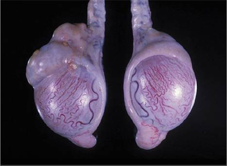
Figure 13.12 Sperm granuloma in the head of the epididymis of the testis on the left, from a polled buck. Source: Courtesy of Dr. M.C. Smith.
Back pressure eventually leads to degeneration and even mineralization of the testicular stroma (Corteel et al. 1969; Soller et al. 1969).
A partially split scrotum has been associated with increased frequency of sperm granulomas in Germany (Schonherr 1956), but appears to be a desirable trait in breeds native to very hot climates, where cooling of the testes is facilitated (Almeida et al. 2010). A sperm granuloma might develop after vasectomy.
Clinical Signs and Diagnosis
Some animals are sterile from the beginning, because all of the efferent ducts are affected. Others are initially fertile, but become infertile if bilateral granulomas form. Normal libido persists.
By the end stage, both the firm granuloma (lentil to egg sized) in the head of the epididymis and the reduced size of the testis are palpable. Less commonly, granulomas form in the body or tail of the epididymis. Normal epididymis is clearly softer than normal testis (Munchen 1951). In ultrasonographic evaluations, appearance of sperm granulomas (in a ram) has been described as fluid-filled structures within a ring of echogenic tissue (Buckrell 1988). Ultrasound examination is also useful for demonstrating testicular mineralization, the end stage of degeneration induced by back pressure.
Correction of this defect is not possible. The diagnosis can be confirmed by gross and histologic examination after castration or slaughter.
Prevention
Because sperm granulomas are very strongly correlated with the homozygous polled condition, breeders can avoid the expense of raising animals that will be (or become) sterile because of this defect by culling all young bucks that are by phenotype homozygous for the polled gene. These bucks have very smooth, well-separated protuberances on the poll. Heterozygous bucks, in contrast, typically have bean-shaped protuberances that converge anteriorly, and many have irregular horny bosses that become evident by 3-6 months of age (Ricordeau et al. 1972a). If polled bucks are kept for breeding, their potential fertility should be monitored regularly by palpation and semen evaluation.
Segmental Aplasia of the Epididymis
In a slaughterhouse study of 100 bucks in Brazil, three were found to have segmental aplasia of the body of the epididymis. One buck was affected bilaterally. Two bucks were horned and one (Moxoto breed) was polled. The head of the epididymis was distended, the body could not be palpated, and the tail was reduced in size (Humenhuk and Vinha 1976). One buck with segmental aplasia was detected in a slaughterhouse survey of 1000 feral male goats from Australia (Tarigan et al. 1990).
Scrotal Hernia
Scrotal and inguinal hernias occasionally have been described in goats (Abdin-Bey and Ramadan 2001). They are seen more commonly in sheep and generally presumed to be hereditary in that species. Clinical signs of a scrotal hernia include distension of one side of the scrotum with a freely movable, fluctuant loop of intestine. The testis may be atrophied. If a hernia is identified in a buck and surgical correction is desired, the buck should be castrated bilaterally.
Other Scrotal Lesions
Wounds (shearing cuts, dog bites, vasectomy) involving the scrotum may result in establishment of a purulent orchitis or periorchitis. Fly strike and grass awn migration might also occur, although these conditions are better described in rams. Some breeders in Texas believe that a split scrotum increases the risk of injury to the scrotum by burrs or brambles (Drummond 1988). Other causes of scrotal dermatitis include mange (Kambarage 1992), bacterial infections, zinc deficiency, and frostbite; the reader is referred to Chapter 2.
Elastosis and fibroblastic proliferation of the intima of the testicular artery have been observed in goats, as has calcification of the wall of the testicular artery and veins (Panebianco et al. 1985). Possible effects of these vascular lesions on fertility are unclear. Varicocele does not seem to have been reported in goats.