The Buck
The male goat is called a buck or billy. Many dairy goat owners are offended by the term “billy,” and Angora breeders are often confused by “buck,” because in many parts of the United States the ram (male sheep) is referred to as a buck.
The buck is frequently described as half of the breeding herd. Many owners find him the more difficult half to control. Because of his size and strength and his distinctive odor, the buck is often skipped when the does are dewormed, vaccinated, given selenium injections, or foot trimmed. Unless pens are massive and fences both sturdy and high, a resident buck breeds does earlier in the breeding season than desired and leaves no written records of his activities. Owners choosing to raise or purchase a buck for breeding would do well to read lay articles on the subject of buck management (Hicks 1987) before getting into this phase of goat husbandry.
Anatomy
The anatomy of the male goat's reproductive tract has been reviewed elsewhere (Smith 1986c; Garrett 1988; Constantinescu 2001). The scrotum is pendulous and in some animals it is divided to a greater or lesser extent into two sacs. There is little evidence that this trait (bipartite scrotum) is detrimental to fertility in the absence of other anatomic defects.
The testes are present in the scrotum at birth and are positioned with the longitudinal axis vertical. Approximately 15-20 efferent tubules collect spermatozoa from the testis and join the head of the epididymis, which is located on the dorsolateral aspect of the testis. The body of the epididymis is caudal and slightly medial, and the tail of the epididymis, where semen is stored before ejaculation, is ventral. Blood is supplied to the testis by the testicular artery, which is precooled by its proximity to the pampiniform venous plexus in the spermatic cord. The cremaster muscles and smooth muscle in the wall of the scrotum also serve to control the testicular temperature by adjusting the position of the testis relative to the abdomen.
The deferent duct passes dorsally on the medial side of the testis and spermatic cord to the inguinal ring, then passes medial to the ureter and widens to form an ampulla, before emptying into the pelvic urethra on the side of the seminal colliculus. The vesicular glands cover the dorsal aspect of the ampullae. Their excretory ducts open at the seminal colliculus, usually through common ejaculatory ducts and orifices that also serve the deferent ducts (Constantinescu 2001). The prostate gland is disseminated in the wall of the pelvic urethra and opens into its lumen by many small ducts. A recess extends caudodorsally from the urethra, where the pelvic urethra turns ventrally after leaving the pelvic canal. The bulbourethral glands open into the lumen of the urethral recess, on the fold of mucous membrane that separates the recess from the pelvic urethra.
The bulb of the penis (corpus spongiosum penis) surrounds the penile urethra and is itself covered by the bul- bospongiosus muscle ventral to the anus. There are two crura of the penis that are formed by the corpus caverno- sum penis surrounded by the heavy tunica albuginea and are covered by the ischiocavernosus muscle, which is important for erection. The hemodynamics of penile erection have been reported by Beckett et al. (1972a, b). Paired retractor penis muscles insert ventrally on the fibroelastic penis just distal to its prominent sigmoid flexure. The penis ends with the glans penis, but the urethra continues as the urethral process, which extends 3 or 4 cm past the glans.
Physiology and Sexual Development of the Male
Breed, age, and nutrition all contribute to the onset of sexual maturity. In Boer goats, spermatogenesis was found to begin as early as 84 days of age, with spermatozoa present in the epididymis at 140 days of age (Skinner 1970). In another study of 20 Boer goats, puberty (as defined by presence of spermatozoa in the ejaculate) was reached at 115-234days (Louw and Joubert 1964). Fast-growing, well-fed kids are able to breed sooner than starved males born at the same time.
Male Boer goats born in the rainy season reach puberty sooner than those born in the dry season in South Africa (Bezerra et al. 2009). In dairy breeds in the United States, many bucklings are fertile by 5 months of age, but successful breeding has occurred as early as 3 months of age. Some other breeds, under other management conditions, mature much later (Elwishy and Elsawaf 1971).Natural adhesions of the urethral process and glans penis to the prepuce render the immature male incapable of copulation (Figure 13.11). Under the influence of testosterone, the urethral process becomes free, beginning at its tip, and finally the penis separates from the preputial mucosa (Skalet et al. 1988). This separation never occurs in the early-castrated wether.
Serum testosterone concentrations in mature bucks in temperate climates vary with the season; the highest are in
the fall breeding season. Increases in testosterone are preceded by increases in luteinizing hormone from the pituitary. Testosterone rises earlier in bucks provided with a high-quality diet than in those that are underfed (Walkden- Brown et al. 1994). Mean values in yearling Pygmy goats in the fall reach 15 ng/mL (Muduuli et al. 1979). In an Australian study of six Angora bucks, the mean androgen level was approximately 10 times more in March (10.25 ng/ mL) than in July, at the end of the breeding season (1.05 ng/ mL) (Ritar 1991). Strong seasonality and breed differences were also demonstrated in four Mediterranean breeds (Todini et al. 2007). There is also a diurnal pattern (Bosu and Barker 1982), although this pattern was not consistent throughout the year in another study (Ritar 1991). In Black
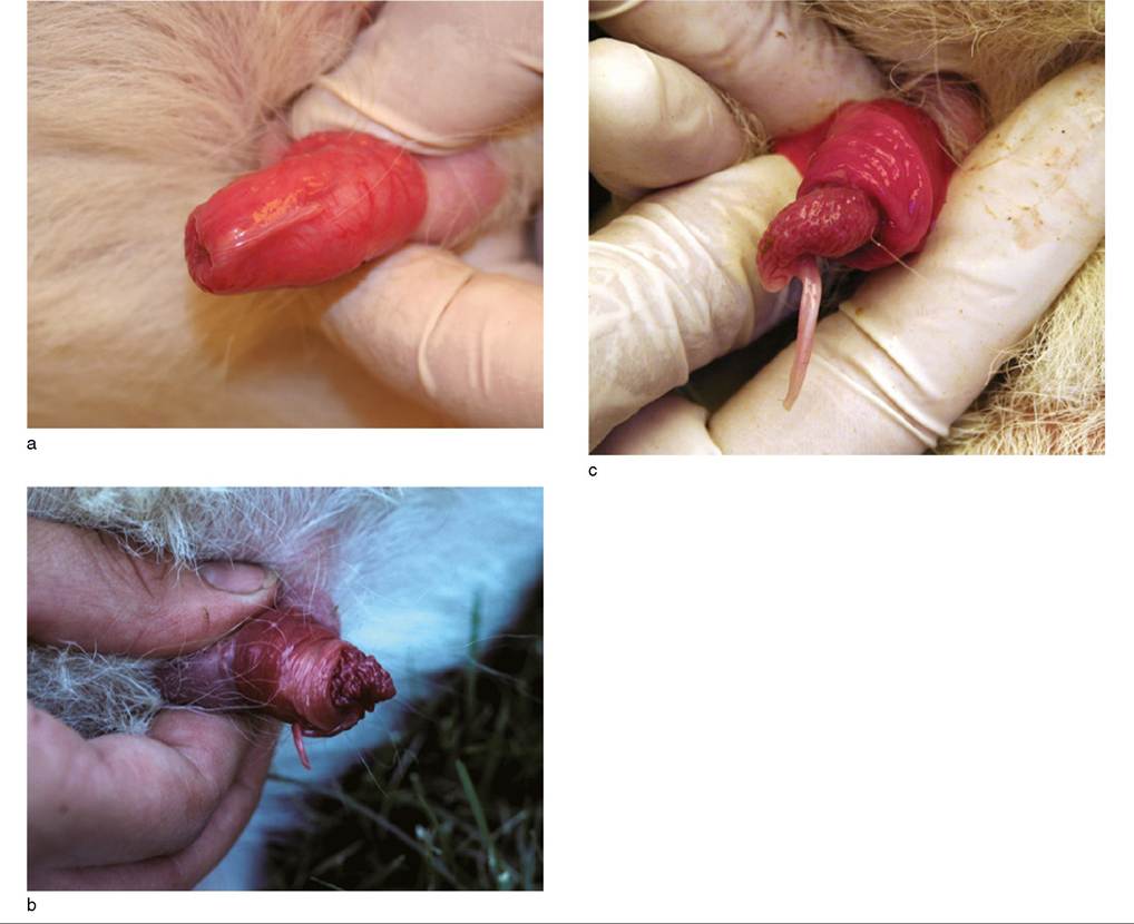
Figure 13.11 Normal maturation of the penis. (a) The urethral process is completely adhered to the lining of the prepuce. (b) Separation of the urethral process and glans penis has begun, giving a striking, although normal, red cauliflower-like appearance.
(c) The urethral process and glans penis are completely separated from the prepuce, and intromission is now possible. Source: Courtesy of Dr. M.C. Smith.Bengal goats, higher testosterone concentrations have been reported in 2-3-month-old kids (7 ng/mL) than in 3-5-month-old (3 ng/mL) or mature bucks (5 ng/mL) (Georgie et al. 1985). When samples are taken for analysis, testosterone appears to be stable in the plasma or serum of bucks for at least 24 hours (Fahmi et al. 1985).
The buck odor, originating in part from sebaceous glands caudomedial to the horns, contributes to the breeding success of a male. The odor stimulates onset of cyclicity, demonstration of overt estrus, and receptivity in females. Bucks that were descented during disbudding may be ignored or rejected by some choosy does. The glands are testosterone responsive and the odor is most intense during the breeding season (Walkden-Brown et al. 1994).
Sexual behavior and sperm production also vary with season. In a French study, the percentage of bucks that failed to ejaculate increased to as high as 25% in the non-breeding season, especially May to August. Testicular weight and total spermatozoa in the ejaculate also decreased out of season (Delgadillo et al. 1991). Similar seasonal variations have been seen in semen quality of dairy bucks in England (Ahmad and Noakes 1996b). Photoperiod control, alternating two months of long, 16hour days with two months of short, 8-hour days, largely abolished the seasonality in sexual activity and semen characteristics (Delgadillo et al. 1991). This could be very useful for managing semen production by bucks at artificial insemination centers.
The effect of rearing conditions other than nutrition on the development of sexual activity in bucks has received little attention. In one study, 20 male goats at weaning were raised in individual pens and an additional 16 male goats were group-housed with 4 females of the same age that had been ovariectomized before puberty.
Collection of semen with an artificial vagina (AV) was begun between 7 and 8 months of age. There was no difference in sexual behavior or sperm output between the two groups, but the average semen volume was significantly higher in the group-housed bucks, caused by increased seminal plasma (Orgeur et al. 1984). A “female effect,” where exposure of bucks to estrous does stimulates pulsatile release of luteinizing hormone by the buck, with consequent increase in testosterone production, has also been noted (Gordon 1997).Breeding Soundness Examination
The ability of the buck to be a successful, highly fertile breeder can never be guaranteed, but close evaluation should permit identification of problems that might interfere with fertility. This is ideally done several months before the intended onset of breeding, so that the buck can be treated or replaced if found to be “unsound.” However, it is more difficult to evaluate libido outside the fall breeding season.
The Society for Theriogenology has developed and revised scoring methods for evaluating the bull and ram. A formal system for the buck has not been widely used. It is inappropriate to assign points for each portion of the examination and then compare the total with an “acceptable” value. Superiority in one aspect of the evaluation does not compensate for poor performance elsewhere. Recently, Tibary et al. (2018) have described a breeding soundness exam for bucks as well as rams.
Physical Examination
The first portion of the breeding soundness examination is a thorough physical examination for general health (Ott 1978; Ott and Memon 1980; Memon et al. 2007; Tibary et al. 2018). Body condition, color of mucous membranes, and sleekness of haircoat should be evaluated for evidence of parasitism, malnutrition, or chronic infection. Teeth, eyes, feet, and joints should be in good condition so that the buck can continue to eat well and can follow and mount the doe in estrus.
The buck should be free of known genetic or possibly hereditary defects such as hernias, jaw malformations, or supernumerary teats.
The presence of extra teats apparently has no relationship to the fertility of the buck (Schonherr 1956), but if the trait is passed to the next generation, the buck’s offspring may be less suitable for milking. Next, the reproductive organs should be examined. This includes palpating the scrotal contents (and ultrasounding if possible) and inspecting the prepuce and penis for various abnormal conditions described in detail in later sections.Scrotal Circumference
Although scrotal circumference or testicular diameter charts are lacking for most breeds of goats, it is reasonable to assume that, as for bulls and rams, the male with the larger testicular size at a given age is likely to produce more sperm. Scrotal circumference is positively correlated with bodyweight and serum testosterone (Bezerra et al. 2009). The risk that the goat has testicular hypoplasia or an intersex condition (polled intersex or freemartin) is also less when scrotal circumference is larger. It has been suggested that the habitual abortion trait in Angoras is associated with small testicular size (Van Heerden 1964).
Some researchers selecting sheep for accelerated lambing and year-round lambing programs believe that rams that show little seasonal (from October to April in northern latitudes) decrease in scrotal circumference sire ewes that have fewer seasonal breeding tendencies (Ringwall et al. 1990). This possibility should be investigated in commercial goat herds striving for out-of-season breeding. Obviously, large and unchanging would be better than small and unchanging. Photoperiod effects appeared to account for a 2 cm difference in scrotal circumference in Alpine and Nubian bucks kept in environmental chambers (Nuti and McWhinney 1987).
The scrotal circumference can be measured with a specially designed metal tape (Lane Manufacturing, Denver, CO, USA) or with any other measuring tape, or even a cord whose length can be compared with a ruler. The circumference is measured at the widest part of the scrotum with both testes held at the same level; it is important not to distract the testes by grasping the neck of the scrotum (Memon et al. 2007). When bucks have similar age and bodyweight, those with above-average scrotal circumference may be expected to produce offspring with earlier sexual maturity and greater fertility. Older bucks are expected to have larger testes and more sperm, but do not underestimate the potential breeding capacity of a young buck.
As an approximate guideline for dairy goat breeds in the United States, bucks weighing more than 40 kg should have a scrotal circumference of at least 25 cm (reviewed by Tibary et al. 2018). Bucks of British breeds had a mean scrotal circumference of 24 cm when they reached sexual maturity at 5.5 months, but the value decreased by several centimeters in the following January and February (Ahmad and Noakes 1996a, b). In another study, the scrotal circumference of mature (3-4-year old) Nubian bucks of proven fertility was 30-33 cm (Skalet et al. 1988). In 10 young bucks of German breeds, the scrotal circumference was 24-28 cm in October, when the bucks were 8 months old and weighed 26-52 kg. Scrotal circumference decreased by a median of 3 cm when poor hay was fed for two months, then increased again by February with improved nutrition (Arbeiter 1963). A study of Creole goats found that nutrition, rather than photoperiod, determined scrotal circumference in this breed as managed locally (De la Vega et al. 2006). Likewise, Australian cashmere bucks on a high-quality diet had markedly higher scrotal circumference than bucks fed a low-quality diet. In both groups the testicular weight dropped through the fall breeding season (when voluntary feed intake was low) and reached a low point early in the winter (Walkden-Brown et al. 1994). In Jordan, Damascus goats had the largest scrotal circumference in the spring, when day length was increasing, which is their normal breeding season (Al-Ghalban et al. 2004). In a young, growing buck, a sudden increase in testicular size indicates the onset of spermatogenesis (Skinner 1970; Bongso et al. 1982).
As an alternative to scrotal circumference, scrotal volume can be measured by measuring the amount of water displaced when the scrotum is immersed. In one study of six Angora bucks, the scrotal volume peaked nine weeks after day length began to shorten (Ritar 1991).
Semen Collection
Collecting and evaluating semen (Eaton and Simmons 1952; Chemineau et al. 1991; Memon et al. 2007) comprise the final portion of the breeding soundness examination. The most representative semen samples for laboratory evaluation are obtained by AV (Barker 1958; Austin et al. 1968). The technique has been described elsewhere (Refsal 1986; Memon et al. 2007). Most bucks of normal libido in the breeding season try to mount any restrained goat, though a cycling doe treated two days previously with 5 mg of prostaglandin F2 alpha or a doe or wether treated with estrogen (1 mg estradiol administered one or two days previously) may make a more willing mount. Assuming that the temperature of the AV is appropriate (40-45 °C; 104-113 °F), even untrained dairy bucks can usually be collected without difficulty. The volume of an ejaculate is typically 0.5-1.5 mL.
Vocalization by the buck, contraction of voluntary muscles during electroejaculation, and the more dilute semen obtained, make electroejaculation less desirable than the use of an AV (Greyling and Grobbelaar 1983; Memon et al. 1986b). It also is impossible to evaluate libido during electroejaculation. This technique, however, may be the only practical way to collect semen from a range buck (O'Brien et al. 1963), and some practitioners routinely sedate the buck before electroejaculation while being careful to avoid inhalation of rumen contents. One report suggested (but certainly did not prove) that a sine-wave electroejaculator resulted in the collection of more sperm with less distress to the buck than a pulse-wave electroejac- ulator (Carter et al. 1990). The animal is initially set up on its hindquarters to extrude the penis, which can be prevented from retracting by wrapping it in gauze clamped in place with a spring paperclip.
Induction of erection and ejaculation in bucks, rams, and bulls by massage of the glans penis within the sheath has been described (Megale 1968). The animal should be standing unrestrained in a quiet environment. After the penis is extruded from the preputial orifice, ejaculation may occur spontaneously (into a funnel) or semen can be collected with an artificial vagina.
Sperm Morphology
Morphology should be carefully evaluated. Previously, sperm abnormalities were designated as primary if they were thought to have originated in the seminiferous epithelium (i.e., abnormal head shape, abnormal acrosome, midpiece abnormalities, strongly coiled tails) or secondary if they were thought to have occurred in the epididymis (i.e., proximal and distal cytoplasmic droplets, tailless heads, single bent tails). Newer work in bulls favors designation of the various abnormalities according to their location in the head, midpiece, or tail instead (Koziol and Armstrong 2018). It is not yet known which morphologic abnormalities are of greatest significance relative to buck fertility. Tibary et al. (2018) have proposed that there should be less than 30% total sperm abnormalities if a buck is to be deemed a satisfactory breeder.
Many veterinarians mix eosin-nigrosin stain with a drop of semen on a slide and quickly draw the mixture out with another slide to create a smear. Evaluation of the air-dried smear with a bright-field microscope allows differentiation of live (appearing white) sperm from dead ones (red because of uptake of eosin), as well as visualization of many morphologic defects. The use of formol saline, or glutaraldehyde fixation (without staining), and a phasecontrast microscope permits detection of more abnormalities, as will greater experience with semen evaluation. It is best to count at least 200 spermatozoa per sample. The number of each type of abnormality should be recorded. If the first sample yields unsatisfactory spermatozoa, a second sample should be collected on the same day (Schonherr 1956). Repeated semen evaluations during several months may be necessary to establish an accurate prognosis. In particular, a variety of febrile diseases may be followed by poor semen quality that lasts for as long as two months.
A high prevalence of morphologic abnormalities may be associated with either sexual immaturity or degenerative changes. Arbeiter (1964) found 15% abnormal sperm in 144 ejaculates from nine healthy German bucks initially 9 months old. In a two-year study of four “yearling” dairy bucks collected multiple times in fall (November) of two successive years, the percent of abnormal acrosomes and of most other sperm abnormalities was decreased the second year (Chandler et al. 1988). In one study involving Nubian bucks, 65% of sperm cells were abnormal at the onset of puberty, but only 12% were abnormal by 8 months of age (Skalet et al. 1988). Misshapen heads, abnormal midpieces (abaxial, double, coiled, bent, looped), and proximal droplets were common in the young bucks. By 8 months of age, only the midpiece abnormalities were common. Dead sperm are more common in the semen of young bucks in spring and summer than in fall and winter (Ahmad and Noakes 1996b).
In older bucks, the season did not influence sperm abnormalities. Likewise, no seasonal effect on sperm morphology was observed in bucks of native breeds in India (Sahni and Roy 1972), although small increases in percentage of abnormal spermatozoa were noted in bucks in Greece in the non-breeding season (Karagiannidis et al. 2000). However, it has been shown that in hot climates, bucks with a divided scrotum (each testis in a separate scrotal pouch) produce sperm with fewer morphologic abnormalities, presumably because of enhanced cooling. The average percentage of sperm abnormalities was 5.4% for six Moxoto bucks with a divided scrotum and 32.9% for four bucks with no scrotal division collected during the dry season (Nunes et al. 1983). The percentage of live sperm was decreased in the dry season compared with the rainy season, and sperm abnormalities were increased in the dry season (Silva and Nunes 1988).
Few reports are available on the influence of disease on semen quality. Detached heads, tightly coiled tails, and thickened midpieces were prominent in semen samples from an 18-month-old infertile LaMancha buck with testicular degeneration (Refsal et al. 1983). Detached heads and acrosomal defects were increased in goat semen after fever was induced by an injection of Freund's complete adjuvant (Yokoki and Akira 1977). Detached heads were also common (70% of the spermatozoa) in the semen of a Nubian buck with seminal vesiculitis diagnosed by histology (Ahmad et al. 1993). Semen changes associated with various infections are described in the discussion of the individual diseases.
Semen Motility and Concentration
Motility has received much attention in the past. Because it is very difficult to optimally maintain conditions such as temperature, it is common for normal semen (expected motility of 70-90%) to have very poor motility when examined under field conditions. Thus, motility is no longer considered to be an important part of the breeding soundness examination, though Tibary et al. (2018) suggest that there should be at least 50% progressively motile spermatozoa in buck semen from a satisfactory breeder. Concentration of spermatozoa in semen of bucks collected by artificial vagina should be estimated. This can be done by hemocytometer techniques or measurement of optical density, after the instrument is calibrated for buck semen. Concentrations of 2.5-3 billion/mL are normal (Eaton and Simmons 1952). Concentration decreases somewhat with more frequent electroejaculation (Oyeyemi et al. 2001).
An approximation of concentration can be determined by spinning the semen for 10 minutes in a microhematocrit centrifuge (Foote 1958). One spermatocrit point represents approximately 200 ? 106 sperm/mL (R.H. Foote, Ithaca, NY, personal communication 1992). Spermatocrits of 25-33 in normal bucks have been observed by the author (MCS). A commercial Unopette® system (Becton- Dickinson, Rutherford, NJ, USA) can also be used to estimate sperm concentration (Memon et al. 2007). The yellow color often seen in the seminal plasma is caused by riboflavin (Mendoza et al. 1989; Ahmad and Noakes 1996b) and does not influence semen quality.
Processing Semen for Artificial Insemination
Undiluted fresh semen can be used for artificial insemination when the buck and does are on the same premises, but usually the semen is extended for short- or long-term storage. Methods of semen preservation and insemination have been described elsewhere (Corteel 1981; Haibel 1986b; Leboeuf et al. 2000; Purdy 2006; Nuti 2007; Cseh et al. 2012). Skim milk-based (such as UHT milk) extenders are often used. High concentrations of egg yolk should not be used in an extender for unwashed semen, because a lipase enzyme secreted by the bulbourethral glands (Chemineau et al. 1999) hydrolyzes egg yolk lecithins to products that are toxic to sperm. When semen is to be kept frozen for many months, washing the spermatozoa to remove most of the seminal plasma before freezing has sometimes improved fertility, especially with breeds or seasons when the original ejaculate volume was small (Corteel 1981; Corteel et al. 1984). Non-breeding season seminal plasma is more detrimental to post-thaw motility than is breeding season seminal plasma (Leboeuf et al. 2000). Photoperiod treatment (alternating two months of long days with two months of short days) alleviates adverse effects of the non-breeding season on postthaw semen quality (Chemineau et al. 1999).
Although fertility was low, cryopreserved epididymal sperm collected from postpubertal bucks at necropsy has been successful in producing a pregnancy (Blash et al. 2000). Laboratory evaluation suggests that viable sperm can be collected for up to 48 hours if the testes are kept refrigerated after removal from the body (Turri et al. 2014).