The Acute Phase Response
Acute inflammatory processes can trigger an acute phase response. Infectious causes are common, but many noninfec- tious causes (e.g., trauma, strenuous exercise) are documented.
The body's response to the inflammatory insult is altered liver production of many proteins, termed acute phase proteins (APPs). A few proteins are negative APPs, and their production decreases with acute inflammation. Albumin is the most prominent negative APP, and levels will decrease in circulation proportionate to its half-life in each species. For example, the albumin half-life is 19.4 days in horses and 16.5 days in cattle, so hypoalbuminemia due to inflammation will take several weeks to develop in these species.6,18 Transferrin and transthyretin (prealbumin) are other negative APPs, as is apolipoprotein A1 (see the Apolipoprotein A1 section later). The presumed role of decreased production of certain proteins is to shift available amino acids to the synthesis of proteins that are useful in host defense.19Most acute phase reactants are positive APPs, meaning production increases with inflammation. These proteins will migrate into the α and β regions on SPE or PPE. The positive APPs are a diverse group, including proteins involved in coagulation, opsonization, iron regulation, and limiting tissue injury. Protein electrophoresis can reveal increased globulin fractions that are composed largely of APPs. Some diseases cause fairly specific alterations; for example, internal parasitism in diarrheic horses caused increased β1 globulin levels that were significantly higher than those in diarrheic horses with other disorders.20 Nonprotein assays are also used to diagnose an acute phase response; for example, serum iron outperformed plasma fibrinogen as an indicator of acute inflammation in horses.21
Measurement of individual APPs can be useful diagnostically and is not limited to blood; assays for APPs in milk and saliva have also proven useful.22-24 The following is a list of major APPs in large animals.
Fibrinogen
Fibrinogen is a useful marker of inflammatory disease in large animals. Increased plasma fibrinogen, termed hyperfibrinogenemia, can occur in a myriad of conditions. Fibrinogen is semiquantitatively measured via the heat precipitation method, along with total plasma protein, as part of the routine complete blood cell count (CBC). Serum samples cannot be used to measure fibrinogen. The heat precipitation method cannot be used to detect hypofibrinogenemia (see Chapter 27).
As for other proteins, fibrinogen concentration can be increased due to dehydration. Calculating the plasma protein/ fibrinogen (PP/F) ratio is useful when fibrinogen is increased. If the PP/F ratio is greater than 15, hyperfibrinogenemia is likely due to dehydration. If the PP/F ratio is less than 10, the patient has true hyperfibrinogenemia (due to inflammation).25 Severe inflammatory disease in horses and ruminants consistently causes hyperfibrinogenemia, although the increase in fibrinogen may not be proportionate to the degree of inflammation. Increased fibrinogen often precedes development of an inflammatory leukogram in ruminants, particularly in cattle.25 Hyperfibrinogenemia can persist for a period of time postoperatively. In one study more than half of horses undergoing subchondral bone cyst removal still had hyperfibrinogenemia 15 days post operation; in another study fibrinogen remained increased through day 11 following laparotomy and other procedures.26,27
In adult horses a number of conditions, including colic, sterile inflammation, surgical intervention, and chronic internal abscessation, are associated with hyperfibrinogenemia
■ BOX 26.3
Causes of Hyperfibrinogenemia in Horses
Abscess (abdominal or other)
Chronic peritonitis Pleuritis
Pneumonia Osteomyelitis Septic arthritis Cholelithiasis
Neoplasia with inflammatory response Vasculitis (equine purpura haemorrhagica) Cellulitis
Gastrointestinal inflammation Salmonellosis
■ BOX 26.4
Causes of Hyperfibrinogenemia in Ruminants
Acute mastitis, especially coliform
Abscess
Traumatic reticuloperitonitis, pericarditis Salmonellosis
Gastrointestinal inflammation
Pyelonephritis Endocarditis
Pleuritis
Pneumonia
Chronic peritonitis
Necrotic rumenitis
Lymphoma (lymphosarcoma)
Septic arthritis
Cellulitis
Omphalophlebitis
Osteomyelitis
(Box 26.3).26,28-30 Vaccination of horses induces an acute phase response including increases in both fibrinogen and serum amyloid A.31 Fibrinogen response in foals is more variable. In healthy neonatal foals fibrinogen levels are initially lower than adult values and then equilibrate within a few weeks of age.32,33 Hyperfibrinogenemia lags behind leukocytosis as an indicator of inflammation in foals.32,34 Fibrinogen was an unreliable marker of foal sepsis in one study,35 although marked hyperfibrinogenemia of 900 to 1500 mg/dL had utility as a diagnostic marker of foal osteomyelitis.36 Fibrinogen may be useful as an indicator of foal pneumonia due to Rhodococcus equi: One study found that hyperfibrinogenemia had greater specificity than leukocytosis, but another reported better diagnostic performance (encompassing both sensitivity and specificity) of leukocytosis over hyperfibrinogenemia.37,38 Late pregnancy correlates with increased fibrinogen concentration in mares.
In one study fibrinogen concentration progressively increased roughly 1 week prior to foaling, peaked at the time of foaling, then gradually decreased but remained increased over control values for several weeks post foaling.39Fibrinogen is a particularly useful indicator of inflammation in cattle and other ruminants, as they develop substantial hyperfibrinogenemia in response to inflammation (Box 26.4).40 A number of inflammatory diseases in cattle are associated with hyperfibrinogenemia, including peritonitis, mastitis, and pneumonia. Lymphoma and uncomplicated displaced abomasum (without peritonitis) did not present with hyperfibrinogenemia.40 Progressive increase in fibrinogen concentration occurs with pregnancy in cattle, with a significant increase just prior to parturition compared to the 2 days prior to calving.41 Fibrinogen is reportedly low to undetectable in neonatal calves.42 One
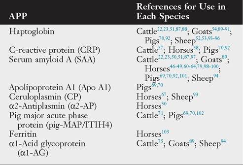
■ TABLE 26.2
Useful Acute Phase Proteins (APPs) in Large Animal Species
study of healthy calves reported a peak in fibrinogen at postnatal day 6 (mean value 400 mg/dL), then a gradual decline until reaching a mean nadir of 250 mg/dL at day 50 followed by a slight rise; values always remained within the reference interval for adult cattle.8 Fibrinogen was significantly increased in calves with suspected septic shock.43