The Identification of the Causative Agent
Despite advanced methods to specifically identify nucleic acids of P. destructans, histopathological investigations remain the gold standard for confirming the disease. The hallmark lesions, i.e.
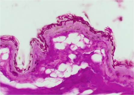
cup-like erosions, have to be documented for diagnosing the disease. The colonisation of only the superficial epidermis without evidence of invasion into deeper tissues (Fig. 13.7) is not accounted for confirming the diagnosis of the disease. Evidence of P. destructans either by swab, touch imprint, culture and/or specific PCR is considered only suspicious of the disease, if confirmation of hallmark lesions by histopathological examination was not pursued.To detect and differentiate P. destructans from closely related fungi in environmental samples, a quantitative dual-probe real-time PCR assay was established based on a single-nucleotide polymorphism (SNP) of the ITS1 region of the genome which was specific to P. destructans (Shuey et al. 2014). As environmental samples
Fig. 13.7 Lightmicroscopy image of superficial colonisation by
Pseudogymnoascus destructans (PAS stain)— these lesions are not diagnostic for WNS
may contain various PCR inhibitors, further investigations were performed to optimise the methodologies. The comparison of different commercial test kits and the addition of a combination of chemical, enzymatic (lyticase and proteinase K) and mechanical procedures for the extraction and purification of nucleic acid from P. destructans in different environmental sample types have led to improved and efficient methods for high-throughput and quantification analyses (Verant et al. 2016).
Conventional PCR tests targeting the internal transcribed spacer (ITS) region of the rRNA gene complex can be used, but they make subsequent sequencing necessary as otherwise cross-reaction with the plethora of P. destructans allies cannot be discriminated (Lorch et al.
2010). A highly sensitive and consistent real-time TaqMan PCR test was developed that detects as little as 3.3 fg of genomic DNA from P. destructans per 25 mL reaction equalling approximately 0.1 genome equivalents. It is designed to target a portion of the multicopy intergenic spacer region of the rRNA gene complex (Muller et al. 2013). This method was employed in a large study to evaluate the fungal load of individual bats by rubbing a swab five times across muzzle and forearm. However, these results require careful interpretation, since Field et al. (2015) compared fungal loads with the amount of cupping erosions detected by histopathology. They found high measures of fungal loads and relatively low numbers of cupping erosions and vice versa, indicating that fungal load and actual tissue damage do not necessarily correlate well.Long-wave ultraviolet (UV) light with a wavelength of 366-385 nm elicits a distinct orange-yellow fluorescence in bat wing membranes that corresponds directly with the fungal cupping erosions in histological sections (Turner et al. 2014) and can be used to visualise epidermal clusters of (invading) fungal hyphae. A non-invasive approach to estimate the severity of P. destructans infection on live bats is based on correlating the fungal load of individual bats assessed via quantitative PCR (Shuey et al. 2014) and the number of foci on wing membranes fluorescing in UV light (Zukal et al. 2016).
13.5