The Interstitial Pneumonias
Amelia R. Woolums
Understanding of the interstitial pneumonias of ruminants has evolved considerably over several decades. Unfortunately, lack of clarity still exists, particularly with regard to terminology.
Terms such as acute bovine pulmonary emphysema, atypical interstitial pneumonia (AIP), fog fever, pulmonary adenomatosis, farmer’s lung, and acute respiratory distress syndrome (ARDS) have been used interchangeably for all of the conditions that follow. This text uses a classification presented by Breeze,1 which places the interstitial pneumonias into four groups: (1) ARDS, (2) hypersensitivity diseases, (3) chronic conditions that may be sequelae of ARDS or hypersensitivity diseases, and (4) parasitic diseases. BRSV infection can also cause AIP; this disease was discussed in relation to the respiratory disease complex of cattle, sheep, and goats in The Bronchopneumonias (Respiratory Disease Complex of Cattle, Sheep, and Goats) section earlier in this chapter.
Acute Respiratory Distress Syndromes
An ARDS is any respiratory condition characterized clinically by a sudden onset of (usually severe) dyspnea (Fig. 31.68) with gross and histopathologic findings consistent with AIP. The characteristic gross findings of AIP are lungs that fail to collapse when the thorax is opened (Color Plate 31.9) and that are heavy and have a firm, rubbery texture on palpation. Interlobular or bullous emphysema is usually present (see Color Plate 31.3), and sometimes the cut surface of the lung has a shiny or wet appearance because of edema. In some cases affected lobules, which are dark red to purple or sometimes grayish, are interspersed with normal-looking lobules, giving the lung a “patchwork” appearance. Although the gross findings are suggestive and often characteristic, other lung diseases can cause similar changes, so a diagnosis of AIP can be confirmed only by histopathologic evaluation.
The histopathologic changes that confirm a diagnosis of AIP include alveolar hyaline membrane formation and fibrin deposition, alveolar and interstitial edema, and type 2 pneumocyte proliferation. Hemorrhage, emphysema, and interstitial inflammatory cell infiltrate can also be seen.The term atypical interstitial pneumonia has often been applied to describe the changes characteristic of AIP, but it has been pointed out that the clinical and pathologic changes in affected cattle are not “atypical” but rather are “typical” of acute lung injury (ALI).1,2 Use of the term acute interstitial pneumonia to describe the characteristic pathologic changes and of the term acute respiratory distress syndrome to describe the clinical picture in an animal with AIP of as-yet undetermined cause is both accurate and all-encompassing. A diagnosis of AIP should be understood as a pathologic diagnosis rather than an etiologic diagnosis because a variety of insults can lead to the same lung lesion of AIP.
■ Definition and Etiology
Acute Bovine Pulmonary Edema and Emphysema
Acute bovine pulmonary edema and emphysema (ABPEE), classically known as fog fever, is an ARDS of adult (over 2 years old) cattle that occurs when these animals are changed from dry, sparse forages to lush, green pastures. It is caused by the conversion of L-tryptophan ingested in the lush forages to a
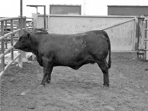
FIG. 31.68 Heifer exhibiting respiratory distress typical of acute respiratory distress syndrome (ARDS). Note the extended head, frothing at mouth, and wide-set front legs. (Courtesy Dr Amelia Woolums, Mississippi State University, Starkville, Miss.)
pneumotoxic compound (3-methylindole [3-MI]), which leads to the development of pulmonary edema, alveolar epithelial hyperplasia, hyaline membranes, and emphysema.
■ Clinical Signs Adult brood cows are most commonly affected because this is the type of animal most likely to be subjected to the abrupt pasture change required to produce the condition.
No breed is resistant. The type of pasture appears to be unimportant, as long as it is lush; ABPEE has been reported on a wide variety of grasses, alfalfa, rape, kale, and turnip tops.3 Signs usually occur within 2 weeks of the pasture change. In severe cases there is an acute onset of very severe dyspnea with a loud expiratory grunt, frothing at the mouth, mouth breathing, and tachypnea. The animals are obviously distressed (as opposed to exhibiting the typical depression that occurs with infectious respiratory diseases) and stand with the head and neck extended and elevated and the nostrils dilated. Temperature and heart rate may be elevated secondary to the severe dyspnea and hypoxia. On auscultation the breath sounds are usually surprisingly soft in view of the gross dyspnea and tachypnea; a few crackles may be heard. Even mild exercise increases the dyspnea and may precipitate collapse and death. As many as 30% of severely affected patients may die, usually within 2 days. Those that survive typically show a dramatic improvement after 3 days. Recovering patients and those that are less severely affected exhibit tachypnea, harsh breath sounds, and crackles and wheezes, particularly in the caudal lung fields. Subcutaneous emphysema may develop. The demeanor of the entire group tends to become more tranquil. In cattle that have repeated episodes of nonfatal ABPEE, a chronic respiratory condition characterized by diffuse pulmonary fibrosis and alveolitis may develop.4 For purposes of differential diagnosis, it is important to note that coughing is not prominent in the individual or the group. The main differential diagnoses are those diseases that cause ARDS in pastured adult cattle, usually in outbreak form. The association of ABPEE with typical management conditions (e.g., changes of pasture) and the absence of coughing, signs of sepsis, and adventitious lung sounds in early cases is also very important. Primary considerations should include the other plant toxici- ties (moldy sweet potatoes, perilla mint, and possibly others) that can be differentiated only by identifying the source. An outbreak of BRSV infection or parasitic bronchitis could cause similar clinical signs and pathology, but these are characterized by more coughing, signs of depression, and more prominent adventitious sounds; fever is common in animals with acute BRSV infection.■ Pathophysiology L-Tryptophan in lush forages is converted by ruminal microorganisms to indole-3-acetic acid and eventually to 3-MI, which is rapidly absorbed from the rumen into the blood. Metabolism of 3-MI by the cytochrome P450 mixed function oxidase system in the nonciliated bronchiolar epithelial (Clara) cells and type 1 pneumocytes results in one or more highly reactive intermediates that bind to intracellular proteins or other macromolecules and thus lead to damage or death of the cells. These intermediates are detoxified by conjugation with glutathione.5 Cellular damage results in degeneration, necrosis, exfoliation of type 1 pneumocytes and Clara cells, and edema. These lesions in turn cause hyaline membrane formation, proliferation of type 2 pneumocytes, and to a lesser extent, proliferation of Clara cells.6 The proliferation of type 2 cells is also known as adenomatosis.
■ Epidemiology As indicated, ABPEE is consistently related to management practices in which hungry adult cattle are suddenly moved from sparse, dry grazing to lush green pastures. The British name “fog fever” arose from the association of the disease with “fog” pastures, which are the lush, green regrowth pastures that occur after hay or silage has been cut. The problem usually occurs in the fall. In the typical pattern in the western United States, cattle are moved from dry summer range onto irrigated or fertilized aftermath pastures. The disease usually appears as a herd outbreak, but individuals may be affected to widely varying degrees7; the morbidity rate commonly approaches 50%, with a case fatality rate as high as 30%.8 Nursing calves typically are not affected.
■ Necropsy Findings In animals that die of ABPEE, ecchymotic to petechial hemorrhages occur in the larynx, trachea, and bronchi, and frothy fluid is present in the airways. The gross and histopathologic lung lesions are consistent with AIP. Congestion, edema, and hyaline membranes cause deep red-to-purple coloration of the cranial lung lobes and a smooth, glistening appearance to the cut surface. Interstitial emphysema with large bullae and gelatinous yellow interlobular edema is common. Histologically, eosinophilic hyaline membranes line alveoli and alveolar ducts, and there edema and proliferation of type 2 alveolar epithelial cells are present. In animals that are killed after 3 to 4 days, emphysema and edema are less obvious, and the lungs tend to be light brown, firm, heavy, and rubbery. There is severe diffuse alveolar epithelial hyperplasia (“adenomatosis”), and large mononuclear cells, multinucleated giant cells, and hyaline membranes are present in alveolar spaces. Edema, eosinophils, and inflammatory cells occupy the septa.
■ Diagnosis Diagnosis is usually presumptive based on the history of recent exposure to lush green forage, typical clinical signs, and gross pathology and histopathology in fatal cases. Measurement of metabolites of 3-MI in blood or urine can be diagnostic, but this test is typically reserved for research applications.9 Thoracic radiographs could be used to identify changes consistent with interstitial pneumonia in valuable individual animals. The results of TTA or BAL cytology have not been reported for cattle with ABPEE, but a nonseptic mixed inflammatory cell response would be expected. There are no unique hematologic or biochemical changes. A stress leukogram may be seen.
■ Treatment and Prevention The stress of handling cattle can precipitate further deaths; because there is no specific treatment for ABPEE, moving cattle to attempt treatment may cause more deaths than it prevents. Some authors maintain that most cases in an outbreak occur within 4 days, that removing the herd from the pasture does not prevent additional cases, and that leaving the herd on the pasture does not result in additional cases; consequently the recommendation has been to handle severely affected cattle only if necessary to remove them to shade or to slaughter.
Others recommend careful removal from the offending pasture. Antihistamines, corticosteroids, epinephrine, atropine, diethylcarbamazine, and diuretics are alleged to be of palliative value, but none of these has been confirmed to be effective in a properly designed trial. Recovery often occurs without therapy in less severe cases. In view of the dangers of handling affected cattle, the questionable efficacy of medical treatment, the probable irreversible nature of severe lesions, and the probability of spontaneous recovery in less severe cases, the best treatment may be no treatment. If treatment must be attempted, affected cattle should be handled very cautiously, and furosemide (0.5 to 1 mg/kg IM or IV once or twice daily) and flunixin meglumine (1.1 to 2.2 mg/kg IV daily or divided twice daily) or steroids (dexamethasone, 0.05 to 0.2 mg/kg IM or IV once daily) may be given. Most fatalities occur in the first 2 days. Severely affected animals that survive may develop chronic emphysema or heart failure secondary to cor pulmonale. Moderate to mild cases often show marked improvement after day 3, with recovery over about 10 days; relapses do not typically occur.Prevention is based on modification of management and prophylactic drugs. Management strategies that have been recommended to prevent the exposure of susceptible cattle to potentially toxic pastures include the following5-8,10:
1. Place the cattle in a dry lot, feed palatable hay for several days, and turn them onto the lush pasture for 2 hours on the first day. Gradually decrease the amount of hay fed and increase the time on pasture over a period of 10 to 12 days.
2. Delay use of lush pastures until after a hard frost.
3. Mow and windrow the pasture before turning cattle out.
4. Use the pasture for young stock (younger than 15 months old) or sheep or other livestock; turn adult cattle out only after the pasture has been thoroughly grazed over.
5. Use the pasture before it becomes particularly lush.
6. Use continuous strip-grazing.
Because such management changes are frequently not feasible, prophylactic medication is a useful alternative. Monensin or lasalocid at 200 mg/head/day PO reduces the conversion of tryptophan to 3-MI.3 Treatment with monensin should be started at least 1 day before pasture change and should be continued for an additional 10 days, whereas lasalocid requires a longer pretreatment period of 6 days.11 Monensin or lasalocid is not expected to be beneficial after the onset of signs. Future possibilities include blockers of the mixed function oxidase system and enhancers of intracellular glutathione levels.
■ Definition and Etiology Feedlot Acute Interstitial Pneumonia
In feedlot cattle an ARDS that is commonly referred to as feedlot AIP occurs. The exact cause of this syndrome is unknown, and it is probably multifactorial. Feedlot management practices typically do not include exposure of cattle to lush forages similar to those that cause ABPEE, and the feeding of moldy sweet potatoes has not been associated with feedlot AIP.12 A variety of causes have been proposed13,14; unfortunately, the amount of research done to confirm or refute the various hypotheses ranges from small to nonexistent. The most commonly suggested possible causes or predisposing factors are (1) feed- associated pneumotoxins such as 3-MI9,15,16 or dietary factors related to protein metabolism,17 (2) chronic bacterial pneu- monia,18,19 (3) gender9,15 and hormonal influences,20,21 (4) chronic small airway injury,18,19,22 (5) BRSV infection,19,23 (6) heat or dust exposure,22,24 and (7) hypersensitivity reactions.22,25,26 These possible causes could also work together in various combinations to cause feedlot AIP.
Support for 3-MI as a cause of feedlot AIP comes from research that showed that levels of a stable metabolite of 3-MI, 3-methyleneindolenine (3-MEIN), were significantly higher in the blood of cattle with AIP than in control cattle.9,15 Increased levels of 3-MEIN may be a result of decreased clearance of the toxin. Loneragan and colleagues also found that 3-MEIN levels were higher in lung tissue of AIP cases as compared with animals without lung disease, but they were not higher than 3-MEIN levels in the lung tissue of cattle with bronchopneumonia; this suggested that 3-MI may contribute to the pathogenesis of both AIP and bronchopneumonia.9 The mechanism by which 3-MI is generated and leads to lung damage was discussed in the Acute Bovine Pulmonary Edema and Emphysema section earlier. If 3-MI does contribute to the pathogenesis of feedlot AIP, it is not clear what about the feedlot diet predisposes cattle to produce high levels of 3-MI. Normal cattle produce some 3-MI through metabolism of dietary proteins. It may be that the protein composition (e.g., the tryptophan concentration) of foodstuffs sometimes added to feedlot rations can lead to spikes in 3-MI production. Cattle with feedlot AIP have been found to have higher ruminal pH values than expected for cattle adapted to a high-concentrate diet. Ruminal pH in AIP cases ranged from 5.6 to 7.2 in one study19 and from 4.9 to 7.4 in another,27 whereas the ruminal pH of cattle adapted to a high-concentrate diet is typically about 5.5 to 5.6.28 Many proteins are relatively basic; therefore the high ruminal pH could be related to abnormal protein metabolism. However, the relatively high ruminal pH could also be caused by anorexia. The concept that abnormal ruminal protein metabolism could contribute to feedlot AIP was also supported by a small study that found increased ammonia levels in the ruminal gas cap of cattle with AIP.29 An interaction between digestive health and AIP was also suggested by the finding that in feedlot pens in which at least one animal had died from digestive disease, the incidence of AIP was approximately 70% greater than in pens in which digestive disease deaths did not occur.17
The hypothesis that bacterial bronchopneumonia contributes to feedlot AIP is supported by the finding by multiple authors that cattle with AIP frequently have superimposed gross and histopathologic lesions consistent with bacterial bronchopneumonia.,, In a study evaluating the bacteria isolated from the lungs of cattle with feedlot AIP that had not received antimicrobial therapy, P. multocida and Mycoplasma spp. were significantly more likely to be isolated from the lungs of cattle with AIP than from the lungs of normal penmates.19 An earlier study found that bacterial respiratory pathogens were not more likely to be isolated from cases of AIP as compared with controls, but the samples in this study were collected at necropsy of animals that received antimicrobial treatment before death in at least some cases.31 It should be noted that no study has yet been able to confirm whether in cases with both lesions the bacterial bronchopneumonia was present before AIP occurred or the bacterial bronchopneumonia developed after the animals had AIP. If bacterial bronchopneumonia leads to the development of AIP, it is not clear how this happens. In humans with ARDS, bacterial infection is often a predisposing factor that appears to lead to ARDS through the induction of high local or systemic levels of proinflammatory cytokines.32 The role of proinflammatory cytokines in cattle with feedlot AIP has not been investigated.
A role for gender and/or hormonal influences in the pathogenesis of feedlot AIP is related to the frequent (although not inevitable)18 finding that the majority of affected animals are heifers.15,17,19 Feeding of melengestrol acetate (MGA), which is fed to heifers to control estrus, has been suggested to contribute to AIP by some authors,20 but others have not found evidence of any association between MGA and AIP.33 Although one research study indicated that MGA could exacerbate experimentally induced AIP in sheep,20 a well-controlled experimental study to determine the effect of MGA on AIP in cattle in the field is needed to clarify the involvement of MGA in AIP. It has also been suggested that growth hormone implants may contribute to development of feedlot AIP, although a survey of feedlot managers did not find data to support this.33
Infection with BRSV was linked to feedlot AIP in an early study.23 Because BRSV infection can sometimes cause AIP, the possibility that a ruminant with lesions of AIP is simply a case of BRSV infection must always be considered. However, cattle with feedlot AIP do not always have a fever,19 which is expected in animals with acute BRSV infection. Moreover, feedlot AIP can occur in outbreaks, but cases often occur sporadically, which would be less consistent with BRSV infection. Neither Sorden and colleagues nor Loneragan and colleagues found BRSV significantly more often in animals with feedlot AIP than in controls.18,31 In another study the difference in frequency of identification of BRSV in feedlot AIP cases as compared with normal penmates approached significance (P =.07).19 Although AIP is by definition “acute,” it is interesting that several authors have reported histopathologic evidence of chronic airway injury (particularly, bronchiolitis fibrosa obliterans) in the lungs of animals with AIP.18,19,22 In one study, the presence of bronchiolitis fibrosa obliterans was identified significantly more often in cattle with AIP than in penmates with no history of treatment for respiratory disease; this was in spite of the fact that AIP cases in the study also had no history of previous treatment for respiratory disease.19 It is not known how past airway injury is related to the development of AIP, although chronic airway injury could be linked to the occurrence of chronic bacterial pneumonia, which has also been associated with feedlot AIP, as described previously. It may be that the chronic airway injury is related to dust exposure, although it is not clear why animals with AIP would be more likely to have airway injury caused by dust exposure than other penmates. Dust exposure has been anecdotally related to AIP,22,24 and feedlot managers often report that efforts to control dust, such as spraying water lightly onto the surface of pens, decreases AIP occurrence when the disease is a problem. However, repeated exposure to feedlot dust or to fungal organisms from feedlot dust did not induce AIP in sheep or goats in experimental studies.34,35 One study of the effect of dust on feedlot respiratory disease found only a weak association between airborne dust particles and respiratory disease.36
High ambient temperatures are thought to contribute to feedlot AIP because several investigators have found that the majority of cases occur in the summer,17,22 but no mechanism by which hot weather might exacerbate AIP has been researched. Hypersensitivity reactions are often suggested to cause AIP. It is true that anaphylaxis can cause pulmonary edema, hemorrhage, and emphysema, with microscopic evidence of hyaline membrane formation.37 However, animals that survive longer than a few hours after an episode of anaphylaxis do not have lung changes consistent with AIP, such as type 2 pneumocytic proliferation and interstitial inflammatory cell infiltrate.38 Therefore an anaphylactic reaction may cause an occasional case of sudden death with lung changes typical of AIP, but anaphylaxis does not explain the majority of feedlot AIP cases. Other types of hypersensitivity-mediated lung disease have pathology that is unlike that seen in feedlot AIP, also making other types of hypersensitivity an unlikely cause of the majority of feedlot AIP cases.1
In summary, the cause of feedlot AIP is unknown, but the strongest support currently exists for some role for (1) factors related to feed, or ruminal metabolism, including 3-MI; (2) infectious respiratory disease, especially chronic bacterial pneumonia and possibly BRSV; (3) gender and/or other hormonal influences; and (4) chronic airway injury, which may be related to infectious respiratory disease. It seems likely that multiple factors can contribute to the development of feedlot AIP, and the factors may act in some as-yet unidentified combination in at least some cases. It is also possible that some causes predominate in some feedlots or individual cases, whereas other causes predominate in other feedlots or individual cases. A study evaluating the occurrence of bacterial respiratory pathogens in cattle with feedlot AIP found bacterial respiratory pathogens in the lungs of the majority of cases in one feedlot and in almost none of the cases in a second feedlot,19 suggesting that bacterial infection played a role in the development of AIP cases in the first feedlot but not the second.
■ Clinical Signs Feedlot cattle with AIP may be found dead in the pen. Clinical presentation includes rapid onset of expiratory dyspnea and tachypnea, although if the respiratory effort is great, the actual respiratory rate may not be greatly elevated. Cattle typically stand with their heads extended and front legs spread apart and exhibit open-mouth breathing (see Fig. 31.68). Frothing from the mouth may also be observed. Rectal temperatures are variable, ranging from normal to elevated.19 Physical examination may reveal cyanosis, tachycardia, and subcutaneous emphysema that extends from the cervical to dorsal thoracic area. Auscultation of lungs reveals dull areas throughout the lungs, along with some crackles. Differential diagnoses of bronchopneumonia, tracheal edema, tracheal obstruction, and hypersensitivity pneumonitis should be considered.
■ Pathogenesis Because the cause of feedlot AIP is unknown, the pathogenesis is also uncertain. If feed-related pneumotoxins cause at least a subset of cases with AIP, the pathogenesis will be similar to that described for ABPEE, 4-ipomeanol toxicity, and perilla ketone toxicity. If bacterial infection is a cause of some cases of feedlot AIP, as is true for some human cases of ARDS, then proinflammatory cytokine production and the resultant inflammatory cascades they initiate are likely involved. More research is necessary to determine the cause and the pathogenesis of feedlot AIP.
■ Epidemiology In the 1999 NAHMS survey of feedlots, AIP was identified as the second leading cause of feedlot mortality, behind bronchopneumonia (shipping fever).39 Mortality rates resulting from AIP of 0.03% to 0.15% have been reported.17,22,30,33 An important feature of AIP is the tendency of the disease to occur most often in cattle on feed more than 60 days,15,19,22,31 as opposed to shipping fever, in which mortality peaks by 45 days after feedlot entry. This means that losses due to AIP deaths are amplified by the fact that relatively more resources in feed and labor have been invested in cattle that die of AIP, as compared with cattle that die of shipping fever. Most cases of feedlot AIP occur in the summer,17,33 but the disease can occur in any season of the year, and most studies report that heifers are disproportionately affected.15,17,19 One survey found that the odds of an animal with AIP being a heifer were 3.1 times greater than the odds of the animal being a steer.17 In feedlot pens in which an animal died from a digestive disorder, the relative risk of AIP occurring was about 1.7, indicating that pens with a digestive disorder death were about 70% more likely to also have an AIP death, as compared with pens with no digestive disorder deaths.
■ Necropsy Findings The gross pathology of feedlot AIP is essentially the same as that described for ABPEE. If there is concurrent bacterial bronchopneumonia, there may be cranioventral consolidation, with fibrin deposition on the pleura, but in cases with no concurrent bacterial pneumonia, the pleura is free of fibrin. Grossly it is common to see a “patchwork” appearance of dark and light-colored lobules interspersed, and the lobules are freely movable (see Color Plate 31.9). Histologically, the most acute cases will have only hyaline membrane formation and alveolar edema, possibly with hemorrhage in the alveoli or interstitium; cases that have been going on longer will have proliferation of type 2 alveolar epithelial cells and inflammatory cell infiltrate into the interstitial space (septa). It is not unusual to find evidence of chronic airway injury, such as peribronchiolar lymphoid cuffing, peribronchiolar vascular fibrosis, and bronchiolitis fibrosa obliterans.18,19 Whether the chronic airway injury is related to the pathogenesis of AIP or is an incidental finding is unknown.
■ Diagnosis Diagnosis of feedlot AIP can be confirmed only by histopathologic evaluation of lung from animals that die or are euthanized because of the disease. Clinical signs and even gross pathology are not definitive; only 65% to 80% of cases identified based on clinical signs were confirmed by histopathologic evaluation to have AIP in two studies.6,15
■ Treatment and Prevention Recommended treatments are similar to those recommended for ABPEE. However, feeding monensin does not preclude development of feedlot AIP, as cattle that die of AIP are often consuming feed including monensin when they contract AIP.15 Treatment is supportive and typically includes administration of antiinflammatory drugs such as steroids (dexamethasone, 0.05 to 0.2 mg/kg once or twice) or flunixin meglumine (1.1 to 2.2 mg/kg IV q24h) and diuretics such as furosemide (1 mg/kg IM or IV q12-24h). Antimicrobial drugs are appropriate given that cases often have superimposed bacterial pneumonia (see Table 31.15). There are no studies evaluating the response of cattle with AIP to any treatment, and such studies would be difficult because there is no perfect method of making an antemortem diagnosis of the disease. However, anecdotal reports indicate that the prognosis is guarded even with treatment. It is important to note that simply moving cattle out of the pen could lead to death from severe respiratory compromise. Because of the risks and uncertainties of treatment and the expected high case fatality rate, immediate salvage slaughter may be the most economic course to take; if salvage slaughter is attempted, remember to observe proper drug withdrawal times.
Because the cause of feedlot AIP is unknown, it is difficult to recommend control measures. Administration of aspirin and of vitamin E have been suggested as rational preventative strategies to counteract inflammatory pathways suspected to be involved in feedlot AIP; however, two trials showed no clear effect of these therapies on levels of 3-MI in treated cattle.40,41 No cattle in these trials developed AIP, so an effect on disease could not be identified. The risk factors identified suggest that management strategies to minimize abrupt dietary changes and to control infectious respiratory disease may be helpful; anecdotal reports also suggest that efforts to control feedlot dust may be helpful.
■ Definition and Etiology
4-Ipomeanol (Moldy Sweet Potato) Toxicity
Moldy sweet potato toxicity is caused by the ingestion of a furanoterpenoid toxin produced by sweet potatoes (Ipomoea batatas) in response to infestation with the fungus Fusarium solani (javanicum). It should be emphasized that this disease is an intoxication and not an allergic response to the fungus.
■ Clinical Signs There is an acute onset of tachypnea, tachycardia, hyperpnea, and dyspnea, with loud expiratory grunting, frothing at the mouth, extension of the head and neck, flaring of the nostrils, and frequent deep coughing. Crackles and harsh bronchial sounds are heard on auscultation. Signs usually occur within 1 day of exposure, and deaths may occur 2 to 5 days later. Differential diagnoses are as for ABPEE (see earlier discussion), which this condition closely resembles, except for the history of exposure and the more prominent cough and adventitious lung sounds.
Pathogenesis When F solani (or closely related species) grows on sweet potatoes, the potato produces several 3-substituted furans, including 4-hydroxymyoporone, which is hepatotoxic. This is converted by the fungus to a series of pneumotoxins, the most abundant of which is 4-ipomeanol. When ingested by cattle in sufficient amounts, this toxin is absorbed, carried to the lungs in the blood, and converted to a highly reactive metabolite by a cytochrome P450-dependent mixed function oxidase system. From this point the pathogenesis is similar to that of ABPEE—that is, the toxin binds to intracellular macromolecules in the cell, causing cellular damage, particularly in Clara cells, type I pneumocytes, and endothelium; edema, hemorrhage, cellular necrosis, hyaline membrane formation, and proliferation of cuboidal epithelium result, with secondary emphysema.
■ Epidemiology The disease usually occurs in outbreak form when groups of cattle are fed damaged sweet potatoes. Morbidity and case fatality rates are high. Calves nursing affected cows are unaffected.42
■ Necropsy Findings The lungs are wet, firm, and large and fail to collapse. Hemorrhages, yellow gelatinous edema fluid, and emphysema with bullae occur throughout. Lobules are dark-red and firm. Microscopic lesions include edema, emphysema, hyaline membranes, hemorrhage, mixed interstitial infiltrates, alveolar epithelial hyperplasia, peribronchiolar fibrosis, and bronchiolitis obliterans.
■ Diagnosis Diagnosis is made based on a history of feeding sweet potatoes and identification of clinical signs and pathology consistent with AIP. Other diagnostic tests as described for ABPEE could also be attempted in valuable individual animals.
■ Treatment and Prevention Treatment has not been investigated. Because the pathophysiologic mechanisms are similar to those of ABPEE, similar recommendations are suggested: handle affected animals with extreme care; if treatment is attempted, furosemide (0.5 to 1 mg/kg IM or IV q12-24h) and flunixin meglumine (1.1 to 2.2 mg/kg IV daily or divided twice daily) or dexamethasone (0.05 to 0.2 mg/kg IV or IM q24h) may be given. The prognosis for moderate to severe cases is grave, regardless of management. Because toxicity is difficult to predict and is usually severe and irreversible when it occurs, the feeding of mold-damaged sweet potatoes should be strictly prevented.
■ Definition and Etiology
Perilla Ketone (Perilla frutescens) Toxicity
Perilla ketone toxicity is an ARDS caused by ingestion of a pneumotoxin found in the leaves and seeds of Perilla frutescens, a common weed in the southeastern United States. This plant is also known as purple mint, perilla mint, wild coleus, and beefsteak plant.43 It is an erect herbaceous annual about 2 m high, with characteristic square stems, an aromatic odor, and opposite, coarsely serrated ovate leaves 5 to 10 cm long and 4 to 8 cm wide with a purplish tint at maturity. The seed and flower stage, which occurs in August to October, appears to be most toxic. The flowers are small, white to purple blooms on a long raceme. The plant prefers semishade, such as damp, open wooded areas.
■ Clinical Signs Animals are often found dead.43 Signs observed include a sudden onset of moderate to severe dyspnea, wheezing, frothing at the mouth, and an expiratory heave or grunt. In less severe cases the cow may pant. Exertion worsens the signs and may precipitate death. Mature cows are most often affected, but deaths have been reported in yearlings and calves. Death occurs in 3 to 7 days in experimental toxicity.43 Differential diagnoses are as for ABPEE, which this condition closely resembles and from which it can be differentiated only by history of exposure.
■ Pathogenesis The volatile oils of P frutescens contain a number of 3-substituted furans that are chemically similar to 4-ipomeanol, the moldy sweet potato toxin. One of these, perilla ketone, predominates in the later growing season (when most toxicities occur) and has been shown to be pneumotoxic when given parenterally to mice, hamsters, goats, calves, and sheep.43 The toxin is absorbed from the rumen, carried to the lungs through the blood, and probably metabolized to the toxic form by the mixed function oxidase system, as for 4-ipomeanol and 3-MI. The pathogenesis from this point parallels that of ABPEE or moldy sweet potato toxicity.
Epidemiology P.frutescens seems to thrive in late summer, when pastures in the southeastern United States are frequently dry and dormant. This also corresponds with the more toxic stage of the plant. Cattle normally avoid the plant when other pasture is available but may be forced to consume it during this critical period. However, under experimental conditions calves were noted to prefer the mint.43 The preseed stage appears to be of relatively low toxicity; the green seed-stage plant is most toxic, especially the seed parts; dried hay from seed-stage plants is less toxic than green plants but is still potentially lethal; and frosted plants appear to have relatively low toxicity.43 The exact toxic dose is unknown, but 2.3 kg of green seed-stage plant and 11.2 kg of hay were lethal for cattle in one trial.43
■ Necropsy Findings The lungs are distended (often bearing the impressions of the ribs), fail to collapse, and are moist, heavy, edematous, and emphysematous. There are often bullae, pleural effusions, and froth in the airways. Histologic characteristics are edema, extensive alveolar epithelial hyperplasia, emphysema, and congestion.
■ Diagnosis Diagnosis is based on history of exposure to perilla mint and characteristic clinical signs and lung pathology. Other diagnostic tests as described for ABPEE could also be attempted in valuable individual animals.
■ Treatment and Control Treatment has not been investigated. On the basis of the similar pathophysiologic mechanisms, the recommendations for ABPEE should be followed. The prognosis for severe cases is grave, regardless of management. Cattle should be provided with sufficient forage that they do not seek out perilla mint; once they have begun to eat it, they should be fenced away from stands of the plant, and other forage should be provided.
Other Toxic Plants
Brassica spp. (rape, kale, turnip tops) are currently regarded as one of the types of pasture that can precipitate 3-MI-associated disease. The possibility that other specific toxins may be identified in these species has not been excluded. Morbidity and mortality rates appear to be much higher on Brassica spp. pastures than on other lush forages.1 The hepatotoxic effects of pyrrolizine alkaloids are well known, but they also cause lung lesions. Lung lesions develop only in animals with chronic liver lesions, and the minimum dose necessary to produce lung lesions is never less than that which is hepatotoxic; therefore signs of liver disease usually predominate. Crotalaria and Trichoderma spp. are the most common offenders; to a lesser extent, Senecio spp. are a cause. Horses, sheep, cattle, and pigs have been affected. Pulmonary lesions include edema, congestion, hemorrhage, proliferation of bronchiolar and alveolar epithelial cells with megalocytosis, and interstitial fibrosis and cellular infiltration. As with 3-MI, 4-ipomeanol, and perilla ketone, the toxicity of the pyrrolizine alkaloids depends on activation by the mixed function oxidase system; in this case, however, the toxin is probably formed in the liver and spills over into the blood to reach the lungs. Vascular endothelium is probably the primary target for injury (versus the Clara cells and type 1 pneumocytes in the other ARDS).
Toxic Gases
Ruminants may be exposed to a variety of toxic gases in the environment. The most important are ammonia, hydrogen sulfide, carbon dioxide, and methane from excreta and respiration; these can be especially important when excreta is collected in pits or tanks. Other gases include nitrogen dioxide from silos; carbon monoxide from machinery exhausts and heaters; zinc oxide from welding of galvanized metal in barns; chlorine, formaldehyde, insecticides, and other fumes from agricultural chemicals and cleaners; and smoke from fires. In most cases concentrations usually remain below overtly toxic levels, and effects are very subtle. Such chronic low-level exposure may result in decreased disease resistance and depression of growth rates. Slightly higher levels of chronic exposure may cause clinically vague syndromes of lethargy, mild dyspnea, anorexia, depressed growth, excessive lacrimation and salivation, low incidence of sudden deaths over weeks or months, and stillbirths. Acute, severe outbreaks usually occur in tightly enclosed facilities and are related to accidents, power outages, agitation or pumping of manure pits, or other combinations of unusual circumstances. Such outbreaks are characterized by an ARDS of variable morbidity and frequently a high case fatality rate.
NITROGEN DIOXIDE. Nitrogen dioxide (NO2) is a yelloworange to brown gas with an acrid odor that is produced by anaerobic fermentation of green plant material. It is a major component of “silo gas.” Acute exposure of farm workers to high concentrations of NO2 causes a respiratory condition known as “silo-filler's disease,” characterized by severe acute edema and congestion and followed by bronchiolitis obliterans and progressive interstitial pulmonary fibrosis. A similar condition has been induced experimentally in cattle,44 and apparent spontaneous field cases have been reported.45,46 Clinical signs in experimental and apparent field cases include cough, tachycardia, tachypnea, respiratory grunting, depression, anorexia, hypogalactia, extension of the head, open-mouth breathing, fever, salivation, lacrimation, and subcutaneous emphysema. Auscultation reveals decreased breath sounds and crackles. The primary differential diagnoses should include other ARDS of housed cattle that occur in outbreak form, especially exposure to other toxic gases (manure pit gases, zinc oxide, chlorine, carbon monoxide), and hypersensitivity pneumonitis from moldy hay. Nitrate toxicity should also be considered. Clinical pathologic evaluation is of limited benefit. Leukocyte counts remained normal in experimental cases; methemoglobin levels increased to a peak at 30 minutes after exposure and returned to normal in 12 to 24 hours.44 The pathophysiologic mechanism probably involves the dissolution of the NO2 in the water of the respiratory tract to form nitric acid. Nitrates and nitrites are also formed; these are irritating, and the nitrites cause methemoglobinemia. Nitrogen dioxide is also an oxidant itself and may contribute directly to the injury. The disease occurs as an outbreak, usually in housed cattle in proximity to a silo chute in a tight or poorly ventilated barn.45,46 Nitrogen dioxide is heavier than air and layers on top of silage or spills out around the bottom of the silo. Corn silage produces more gas than hay, and a high nitrate content increases the danger. The levels are highest in the first 48 hours after filling the silo but may remain dangerous for 2 to 3 weeks.
Necropsy findings in experimental disease include hyperemia of the upper airways; hemorrhages, fibrinous membranes, and froth in the trachea; distended, noncollapsing lungs with rib imprints; a mottled appearance caused by consolidated lobules alternating with emphysematous lobules; and bullae. Microscopic lesions include alveolar epithelial hyperplasia, large foamy alveolar macrophages, hyaline membranes, hyperemia, hemorrhage, and edema.
Treatment involves the establishment of adequate ventilation; cows should be completely removed from closed buildings if necessary. Corticosteroids have apparently been beneficial in field cases, but no controlled studies have been performed to confirm efficacy. Because of the obvious differences in pathophysiologic characteristics, it would be unwise to extrapolate treatment regimens from those of ABPEE. Suggested empiric therapy might include corticosteroids (dexamethasone, 0.05 to 0.2 mg/kg IM or IV daily), furosemide (0.5 to 1.1 mg/kg IM or IV daily or twice daily), and appropriate antibiotics to prevent secondary bacterial infections.
ZINC OXIDE. Zinc oxide (ZnO) fumes have been associated with an ARDS in cattle.47 Oxyacetylene cutting or arc welding of galvanized pipe results in production of white fumes of zinc oxide containing colloidal particles 0.3 to 0.4 mm in diameter, which can reach the terminal alveoli when inhaled. Construction activities in closed barns containing animals may result in toxicity in animals in close proximity to or in the path of ventilation. All ages may be affected.
Clinical signs in severe cases include acute onset of anorexia, frothing at the mouth, anxiety, extension of the head and neck, mouth breathing, expiratory grunting, tachypnea, tachycardia, mild fever, subcutaneous emphysema, and crackles on auscultation of the lungs. Death may occur within 12 hours. Less severely affected animals exhibit depression, mild fever, and tachypnea. Differential diagnoses include other ARDS of housed cattle that occur in outbreak form, such as exposure to other toxic gases (nitrogen dioxide, manure pit gases) or hypersensitivity pneumonitis. The pathophysiologic process presumably involves direct damage to cells by the ZnO and its products dissolved in the fluid lining the respiratory tract. Necropsy findings include purulent conjunctivitis; subcutaneous emphysema; congestion of the airways; tracheal hemorrhages; stiff, noncollapsing lungs; and pulmonary congestion, edema, and emphysema with bullae. Histologic lesions include pulmonary congestion, emphysema, edema, and mixed cellular infiltrates with a prominent eosinophil component. Treatment of severe cases with epinephrine, antihistamine, atropine, and corticosteroids had no effect in one outbreak, whereas mild cases recover spontaneously. Suggested empiric therapy could include ventilation of the area, dexamethasone (0.05 to 0.2 mg/kg IM or IV daily), furosemide (0.5 to 1.1 mg/kg IM or IV daily or twice), and appropriate antibiotics to control secondary bacterial invaders.
CHLORINE. Chlorine is a greenish-yellow gas widely used in manufacturing. Animal exposure is usually the result of industrial accidents. Exposed animals may be found dead. Signs include depression, profuse nasal discharge, lacrimation, and dyspnea; crackles may be heard on auscultation.48 The toxic effects are the result of the formation of hydrochloric and hypochlorous acids; the latter breaks down to hydrochloric acid and oxygen, both of which are toxic to tissues. Necropsy findings include congestion of the nasal mucosa, tracheitis, and pulmonary edema, hemorrhage, and emphysema. Histologic lung lesions include edema, emphysema, hemorrhage, atelectasis, hyaline membrane formation, and lymphocytic bronchitis. Treatment is empiric; suggestions include corticosteroids (dexamethasone, 0.05 to 0.2 mg/kg IM or IV daily), furosemide (0.5 to 1.1 mg/ kg IM or IV daily to twice daily), and appropriate antibiotics to prevent secondary bacterial infection.
MANURE GASES. Manure gases include mixtures of hydrogen sulfide, ammonia, carbon dioxide, methane, and carbon monoxide.1 Accumulation of these gases can result in asphyxiation of animals in enclosed barns over manure pits.
SMOKE INHALATION. Smoke inhalation injury may occur in animals that survive barn fires. Many clinical changes may not become evident for 24 to 48 hours after the fire. It is important to assess the degree of damage as early as possible and to attempt to anticipate sequelae so that early aggressive therapy can be instituted.49 Common problems to check for include oral burns, conjunctivitis, and laryngospasm. Hoarseness, expiratory wheezes, and carbonaceous sputum indicate potentially serious sequelae. Crackles and wheezes on auscultation may occur very early or may be delayed for hours. Cough, stridor, and tachypnea may also occur. Bright red mucous membranes may indicate carbon monoxide poisoning or burns and may mask cyanosis. Carboxyhemoglobin determinations on iced venous blood (often available at human hospitals), serial arterial blood gas determinations, transtracheal wash, and bronchoscopy are useful in delineating the extent of damage and prognosis.
The pathophysiology of smoke inhalation is complex and involves two main mechanisms: carbon monoxide toxicity and smoke toxicity. Carbon monoxide toxicity results in tissue hypoxia in all organs, especially the brain; oxygen consumption in the fire and the pulmonary effects of smoke may aggravate this hypoxia. Heat damage in animals is usually limited to the upper airways. Smoke toxicity is related to the inhalation of soot, superheated particles, and a variety of noxious gases (e.g., aldehydes, oxides of sulfur and nitrogen, benzene from plastics), which results in the formation of dissolved acids, alkaloids, and other direct irritants in pulmonary fluids. These mechanisms eventually (usually 2 to 24 hours after inhalation) result in alveolar damage, interstitial edema, hypoxia, and secondary bronchopneumonia.
Treatment involves establishing a patent airway with intubation or tracheostomy if necessary. Oxygen, up to 100% for short periods, is indicated; care must be exercised because 100% oxygen can also cause pulmonary damage. IV fluids should be given, with careful monitoring for pulmonary edema. The use of corticosteroids is controversial. Antibiotics are indicated to prevent secondary bronchopneumonia. Bronchodilators such as aminophylline at 6 to 10 mg/kg IV or PO three times daily may help relieve soot-induced bronchospasm.
Hypersensitivity Pneumonitis
■ Definition and Etiology Hypersensitivity pneumonitis, also known as extrinsic allergic alveolitis (EAA) or “bovine farmer's lung,” is an allergic respiratory disease caused by inhalation of organic dusts. Several such conditions are recognized in humans, differing only in the nature of the antigen and the circumstances under which exposure occurs. The classic example, of which the bovine disease is probably the counterpart, is “farmer's lung.” EAA is caused by exposure to the dust from moldy hay, grain, or other plant matter containing spores and products of thermophilic actinomycetes such as S. rectivirgula (formerly M. faeni) and T. vulgarisr°o-0 Other unidentified forms of hypersensitivity pneumonitis probably occur in cattle.
■ Clinical Signs EAA is a disease of confined adult cattle; consequently it is more common in dairy breeds than in beef breeds. Typically a succession of acute cases occurs during the winter housing period, so the clinician is presented with a group problem in which animals are in varying stages of the disease. The acute form is indicative of recent exposure and is characterized by a sudden onset of dullness, decreased appetite, hypogalactia, coughing, expiratory tachypnea, dyspnea, and cranial-ventral crackles on auscultation of the lungs.50-52 There is a moderate transient fever, which is frequently missed. The chronic form is insidious at onset and may not be detected until there is considerable fibrosis. Animals with the chronic disease may have acute exacerbations as a result of heavy antigen exposure; some may not be detected until turned out in the spring, when increased exercise causes an acute crisis. There is a history of weight loss and coughing for several winters, with remission in the grazing season. Chronic signs include hypogalactia, weight loss, productive coughing, tachypnea, obvious hyperpnea, and widespread crackles and wheezes on auscultation of the lungs, especially in the rostral-ventral areas. Differential diagnoses should include respiratory diseases affecting groups of housed adult cattle in winter. Infectious diseases (viral and bacterial pneumonias) can usually be differentiated from farmer's lung by careful evaluation of factors such as clinical signs of fever and pulmonary consolidation. Therefore the main differential diagnoses are the toxic gases. It should be emphasized that hypersensitivity pneumonitis is not considered an ARDS and that no evidence exists for the involvement of hypersensitivity in the pathogenesis of the ARDS described previously. However, severe cases of EAA with prominent dyspnea and moderate, nonfatal cases of gas intoxication may appear clinically similar. If the group outbreak aspect of EAA is ignored, the clinician who examines one individual may be unable to distinguish this condition from fibrosing alveolitis.
■ Pathogenesis The spores of the thermophilic actinomycetes are 0.7 to 1.3 mm in diameter and can easily reach the alveoli, where they induce both humoral (precipitating) and cellular immune responses. It is thought that repeated exposure results in the activation of a number of immunologically specific and nonspecific cellular and humoral effector mechanisms at the alveolar level, which results in tissue damage.
■ Epidemiology EAA is a problem in areas with wet summers and severe winters, a situation characterized by the combination of moldy hay and housing of cattle in winter. In North America, EAA occurs primarily in the Great Lakes region of the United States and the eastern provinces of Canada.3 Baling and stacking of hay with a high moisture content (over 30%) results in overheating of the stacks. Thermophilic molds become dominant and produce billions of spores, which are released when the hay is distributed for feeding. A similar situation can occur in stored grains. Affected hay is usually dry, friable, discolored, and dusty; however, it is not necessary for hay to be grossly dusty and poor in quality to release large numbers of spores.
■ Necropsy Findings In acute cases the lungs are superficially grossly normal. Closer inspection reveals small gray spots in many lobules, which represent interstitial and peribronchiolar accumulations of lymphocytes. Other lobules exhibit dark red centers of atelectasis surrounded by pale pink raised edges of trapped air that are caused by narrowing of airways with lymphocytic infiltrates. Histologic evaluation confirms the presence of these lymphocytic infiltrates and aggregations, as well as epithelial granulomas, bronchiolitis, and bronchiolitis obliterans. The gross appearance in chronic cases is similar, with the addition of focal areas of interalveolar fibrosis and epithelial hyperplasia; epithelioid granulomas are absent unless a recent acute exposure has occurred.
■ Diagnosis Precipitating antibodies to M. faeni are found in the serum in most cases. However, two precautions are necessary. First, the presence of precipitating antibodies only indicates exposure to the antigen; many normal animals also have antibodies. Second, it is possible that other as-yet unidentified antigens may cause allergic respiratory disease in cattle. The presence or absence of titers is therefore not necessarily diagnostic of the clinical status of the animal, and the magnitude of the titer does not reflect the extent of disease.
■ Treatment and Prevention Treatment and control center around removing the offending antigen, which is frequently difficult from an economic and management standpoint. Corticosteroids (dexamethasone, 0.05 to 0.2 mg/kg IV or IM daily) may be beneficial in acute cases. Suggestions to decrease the molding of hay or degree of exposure include making silage instead of hay, ensuring proper drying before baling, and feeding hay outside. If the condition can be arrested before significant fibrosis occurs, the prognosis is good.
Anaphylaxis
In ruminants the lung is the major target organ in immediate (type I) hypersensitivity reactions. Precipitating antigens include vaccines, drugs, blood, Hypoderma bovis or Hypoderma Iineatum larvae, insect bites, and bee stings. Signs of anaphylaxis usually develop in 10 to 20 minutes and include severe acute dyspnea, with flaring of the nostrils, extension of the head and neck, open-mouth breathing, frothing of the mouth, hyperpnea, and abduction of the elbows. Pharyngeal and laryngeal edema cause stertor and inspiratory dyspnea. Urticaria may occur in some cases, such as milk allergy in Jersey cows. Shivering, salivation, lacrimation, pruritus, diarrhea, fever, edema (eyes, muzzle, anus, and vulva), collapse, nystagmus, cyanosis, and discharge of froth from the nostrils also may occur. On auscultation there are harsh breath sounds, large airway sounds, and crackles. The primary differential diagnosis is peracute interstitial pneumonia. The pathogenesis involves an initial exposure to the antigen, which results in a genetically determined production of IgE. This antibody attaches to receptors on mast cells and basophils. On subsequent exposure to the antigen, bridging of the Fab parts of the antibody on these cells results in degranulation, with release of a variety of mediators such as histamine, bradykinin, 5-hydroxytryptamine, serotonin, slow- reacting substance of anaphylaxis (SRS-A), eosinophil chemotactic factor A, platelet activating factor, kinins, and prostaglandins. These mediators result in a cascade of vascular events, notably increased vascular permeability, which in turn results in acute severe edema. Pulmonary venous constriction, pulmonary artery hypertension, splanchnic pooling, increased mucus secretion, and bronchospasm also occur. Ruminants that die of anaphylaxis have severe pulmonary congestion and edema, laryngeal edema, and froth in the airways. Lung lesions of ARDS may be present.
The treatment of choice is epinephrine, 4 to 8 mg (4 to 8 mL of 1: 1000 solution) IV or SC or 1 to 5 mg IV for an average 500-kg cow and 1 to 3 mg IV or SC for an average adult sheep or goat. Epinephrine has a short half-life, and animals should be observed closely for relapse. Ancillary therapy includes corticosteroids (dexamethasone, 0.1 mg/kg IM or IV; prednisolone, 2.2 mg/kg IM or IV daily). Other supportive therapies include shock doses of IV fluids (40 mL/kg/h), aminophylline, diuretics such as furosemide, oxygen therapy, and tracheostomy if necessary.
Miscellaneous Chronic Pneumonias
At least two chronic interstitial pneumonias of uncertain cause have been identified in cattle: fibrosing alveolitis and bronchiolitis obliterans. These are typically chronic diseases of individual animals.
Fibrosing Alveolitis
Fibrosing alveolitis is by definition a chronic disease of unknown and possibly multiple causes, characterized by diffuse inflammation of the lung beyond the terminal bronchiole. Approximately 50% of cases produce positive results for precipitating antibody to M. faeni, and it is possible that these cases represent chronic farmer's lung, whereas others (that have negative M. faeni results) may be the chronic stage of hypersensitivity to other unidentified antigens.1 Field investigations have failed to substantiate any connection between fibrosing alveolitis and repeated episodes of ARDS such as ABPEE. Furthermore, recovery from a single dose of 3-MI does not result in lesions typical of fibrosing alveolitis. However, repeated doses of 3-MI at weekly intervals experimentally cause fibrosing alveolitis-like lesions. Fibrosing alveolitis occurs in individual adult cattle (usually over 6 years of age) in both housing and pasture conditions. The history is usually that of a chronic progressive respiratory disease several weeks to 2 years in duration. Affected cattle remain bright and alert and continue to eat until the terminal stages of cor pulmonale and heart failure intervene. Signs include marked weight loss; consistent coughing; tachypnea; very marked hyperpnea, even at rest; and dyspnea after mild exertion. Auscultation may reveal crackles in the rostral-ventral lung field and widespread wheezes. Fever is not apparent. The primary differential diagnoses to consider are other chronic respiratory conditions of individual adult cattle. Chronic suppurative pneumonia and metastatic pneumonia can usually be differentiated from fibrosing alveolitis by careful physical examination and detection of depression, anorexia, shallow respiration, thoracic pain, and hemoptysis. ARDS caused by feed-associated pneumotoxins and lungworms are typically group problems, with at least some severe cases in the group. Differentiation from EAA may be impossible if the group outbreak nature of farmer's lung is not apparent; in fact, at least some cases of fibrosing alveolitis may represent chronic farmer's lung. At necropsy the lungs are very pale, firm, and heavy throughout. Scattered lobules are gray-red, slightly collapsed, and edematous. Thick mucus may be found in the airways, and there is frequently right ventricular hypertrophy as a result of cor pulmonale. Histologic changes are diffuse and include interalveolar fibrosis and infiltration with plasma cells, lymphocytes, mast cells, and interstitial cells; obliteration of alveolar spaces; mononuclear exudates in alveoli; alveolar epithelial hyperplasia; bronchitis; and bronchiolitis. The epithelioid granulomas characteristic of acute farmer's lung are absent. No treatment exists, and the lesions are irreversible.
Bronchiolitis Obliterans
Bronchiolitis obliterans is a chronic respiratory condition of yearling or young adult cattle characterized by a deep infrequent cough, tachypnea, hyperpnea, and an exaggerated expiratory effort. There is no fever. The cause is unknown; the condition is speculated to be a sequela to viruses (BRSV PIV-3, IBR), parasites such as Dictyocaulus viviparus, or hypersensitivity pneumonitis. The lungs appear grossly normal at necropsy, except that they do not collapse. Histologic examination reveals extensive bronchiolitis and bronchiolitis obliterans, with epithelium-covered polyps with a connective tissue core projecting into and obstructing the lumen.1