Urinary Tract Infection
Brandon J. Dominguez
UTI in ruminants typically present as cystitis, ureteritis, and pyelonephritis from ascending infection with C. renale or E. coli.1 Other coliform species2 and close relatives of C.
renale may be involved with lesser frequency.3 Less commonly, Salmonella spp., T. pyogenes, P. aeruginosa, Proteus spp., Streptococcus spp., and Staphylococcus spp., among others, have been implicated in renal infections of hematogenous origin (suppurative embolic nephritis).4-7 In small ruminants, Corynebacterium pseudotuberculosis has also been cultured from renal infections.8Clinical Signs
Cystitis is suggested clinically by dysuria and pollakiuria; hematuria and pyuria may not be grossly apparent. Affected animals may dribble urine, tread their feet, swish their tail, or retain an arched stance after voiding. Occasionally, blood, purulent debris, or urinary crystals may be found on the preputial tuft or ventral commissure of the vulva.1 The bladder may be thickened and painful on rectal palpation, and on cystoscopy, catarrhal, hemorrhagic, fibrinous-purulent, and fibrinous-hemorrhagic lesions may be observed.9 Generalized signs of malaise are not seen if infection is limited to the bladder.1
Acute cases of pyelonephritis often present with a history of sudden reductions in feed intake and milk production. Fever, tachycardia, tachypnea, decreased skin turgor, enophthalmos, ruminal stasis, scleral injection, and occasional episodes of mild colic may be noticed in addition to signs of cystitis.1 When the left kidney is involved, renal enlargement, pain, and loss of the normal lobulation may be noted on rectal palpation. Affected animals may dribble urine, resulting in phosphate calculi present on the vulvar or preputial hairs.
This may be accompanied by wet hair on the perineum and hind legs, progressing to excoriations due to urine scald.Evaluation of the right kidney in cattle can be accomplished via ultrasound with a 5.0-MHz or lower frequency transducer in the 12 th intercostal space and right paralumbar fossa.10-12 In small or thin cattle, the left kidney may be visible in the left paralumbar fossa on transcutaneous ultrasound. Alternatively, the left kidney can be assessed transrectally with a 5-MHz or higher transducer. In small ruminants and camelids, both kidneys are often visible via transabdominal ultrasound from the right paralumbar fossa. Renal enlargement, abnormal kidney shape, dilated renal calyces, and flocculent echogenic material in the renal pelvis and ureter are suggestive of pyelonephritis.10-12 The ureters may be palpated per vaginam in cattle, with the examiner using their fingers to detect one or both enlarged, painful ureters adjacent to the ventral vaginal wall.1
Chronic pyelonephritis presents with vague signs.1 Weight loss, muscle wasting, poor growth, anorexia, diarrhea, and reduced milk production may be evident from history. No abnormal behavior or posturing during urination may be noted. Pale mucous membranes (due to anemia) may be noted during physical examination. A loss of lobulation on the left kidney may be evident on rectal palpation in cattle; pain and enlargement may not be noted.1 Cystic lesions may be noted ultrasonographically in chronic renal disease.12
Differential Diagnosis
Mild colic may be a symptom of numerous GI or intraabdominal disorders, and urinalysis findings will be normal. Dysuria may be the result of vaginitis, perivaginal abscess, pelvic entrapment of the bladder, vulvar trauma, or postparturient swelling of the vagina and vulva. Urolithiasis can cause dysuria and hematuria, but males are typically affected, whereas females are predisposed to UTI. On rectal palpation in cattle, bladder distention will be evident in cases of urolithiasis, a finding not expected with UTI.
A careful neurologic examination will help determine whether there is underlying bladder paresis or incomplete voiding from neurologic diseases. Hematuria may be due to bleeding from parturition, papillomas in the urinary tract, postparturient hemoglobinuria, or enzootic hematuria. In cases of enzootic hematuria, multiple animals are typically affected and the presence of bracken fern can be demonstrated. In addition, a profound anemia is expected with enzootic hematuria, and if pyuria or bacteriuria is present, it will be mild. Papilloma of the bladder may be ruled out with cystoscopy.Clinical Pathology
A neutrophilic leukocytosis with hyperfibrinogenemia is expected in cases of pyelonephritis. If the duration of the infection has been several days, hyperglobulinemia will develop. Continued, severe proteinuria, resulting in hypoalbuminemia, can lead to diarrhea because of decreased plasma oncotic pressure. As the chronicity of the disease lengthens, anemia may develop. This may be due in part to the decreased production of erythropoietin in the affected renal tissue and from blood loss through urination.1 Measurement of serum BUN and creatinine is critical for prognosis in cases of pyelonephri- tis.9,13 When azotemia and isosthenuria are combined in cases of pyelonephritis, bilateral kidney involvement is indicated and the prognosis for successful treatment is poor.1 Prerenal causes should be considered when evaluating the severity of azotemia.
Urinalysis is necessary for definitive diagnosis of UTI. A midstream or endstream caught sample should provide the most accurate results as contaminants in the distal urogenital tract are purged. Concomitant metritis, vaginitis, or prostatitis may contaminate urine with blood, bacteria, and inflammatory cells. Hematuria, proteinuria, and bacteria should be present in the urine of a UTI-afflicted animal. Specific gravity may be in the range of 1.005 to 1.020.12 A Gram stain of the urine may yield preliminary identification of the causative organism.
Pathophysiology
Initiation of an ascending infection is dependent on the amount and virulence of bacteria introduced into the urinary tract. Urogenital trauma during parturition or obstetric manipulation, abnormal vulvar confirmation, conditions leading to urine retention, and bladder catheterization may provide routes of introduction for pathogens. Impaired bladder emptying due to bladder adhesions, urachal remnant infection, or diseases of the spinal cord may create an environment for the establishment of cystitis and ascending infection. Urethral trauma from catheterization, urolithiasis, breeding, or urogenital papilloma may be adequate to initiate infection.14 Vesiculoureteral reflux due to decreased bladder contractility or ectopic ureters may occur, aiding in the extension of infection up the urinary tract. Ureteral and renal obstruction due to inflammatory debris, hemorrhage, and fibrin deposition may lead to episodic signs of colic. After pyelonephritis is established, necrosis of papillary and tubular epithelium leads to an accumulation of necrotic debris in the renal pelvis, loss of functional nephrons, abscessation, fibrosis, and distortion of the renal shape. Struvite uroliths and other renal calculi may develop when necrotic debris serve as foci of crystal deposition in the alkaline microenvironment created by bacterial urease activity.4
C. renale is a large, pleomorphic, nonmotile, gram-positive bacillus.3 It is grouped with and closely related to two other Corynebacterium that may cause pyelonephritis: C. cystitidis and C. pilosum.5 C. renale pyelonephritis has been documented in a sheep14 and induced experimentally in goats.15 Evidence suggests that C. renale may be maintained in the environment for several weeks.3 The bacterium may be transmitted by direct vulvar contact between females or by splashing of urine from infected animals. Iatrogenic transmission may occur via contaminated obstetrical instruments or urinary catheters.
Males may spread C. cystitidis and C. renale through sexual contact.16 C. renale adhere to the urinary tract epithelium via pH-mediated pili.17 Adherence is enhanced in alkaline conditions and reduced in acidic conditions.18 This pH influence may explain the improvement in clinical signs realized after the feeding of urine-acidifying salts. The organism uses ureolysis and ammonium production to maintain urine alkalinity and promote colonization of the epithelium. A serum antibody response will develop after renal infection. This humoral response is rarely curative, nor does it provide resistance to reinfection.1E. coli, among other coliforms, is frequently implicated in ruminant UTI.1 The serotype(s) and virulence factors of E. coli specifically involved in bovine pyelonephritis are unknown at this time. Because of the ubiquitous nature of this gramnegative coliform, it is thought that infection originates from fecal contamination or a loss of natural defenses in the urogenital tract.
Epidemiology
On-farm prevalence of pyelonephritis in cattle varied from 0.3% to 2.7% in an Israeli study.13 An Iranian abattoir study found a 13% prevalence of urine infected with pathogenic organisms.6 Female animals have a greater potential for infection owing to a shorter urethra in addition to potential contamination and trauma during parturition and breeding. A majority of pyelonephritis cases in cows develop within 90 days post calving, which suggests that the postpartum period is a significant risk period in the development of infection.4 Reproductive tract abnormalities such as pneumovagina, metritis, and poor perineal conformation were identified in 7 out of 15 cows diagnosed with pyelonephritis.1
C. renale has typically been regarded as the most common pathogen in bovine pyelonephritis.2,12 In some studies, E. coli has been found as the more prevalent bacteria in pyelonephritis cases.4,5,19 C.
renale and C. pilosum have been found in healthy cows, whereas C. cystitidis is considered the most virulent pathogen in the C. renale group.5 Once established, C. renale can be difficult to eradicate on a herd level.Necropsy Findings
The bladder will have evidence of hemorrhage, ulceration, and fibrin deposition on the epithelial surface, which may extend through the urethra. Polypoid growths may develop with chronic infection. As these resemble tumors, histopathologic examination is necessary for definitive diagnosis.2 Ureters may be noticeably enlarged on one or both sides and contain purulent debris. Acute to subacute pyelonephritis cases may show gross enlargement of the affected kidney(s). A viscous gray, odorless exudate may be found in the renal pelvis and extend into the medulla and cortex, especially on sagittal section. A Gram stain of the exudate is helpful for differentiation of the cause. Chronic cases of pyelonephritis may have gross distortion of renal size and shape (Fig. 34.19).
Treatment and Prognosis
Aggressive and long-term antibiotics are required for successful treatment of UTI. Diuresis with oral or parenteral fluid therapy is beneficial in facilitating the removal of necrotic debris and bacteria from the urinary tract.1 Nephrectomy in cases of
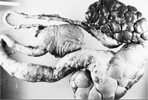
FIG. 34.19 Postmortem specimen of unilateral pyelonephritis and ureteritis in a cow. The affected kidney and ureter are greatly enlarged.
unilateral pyelonephritis that are unresponsive to medical therapy is an option.12 Studies have indicated that there is no apparent limitation on growth and production following unilateral nephrectomy if the remaining kidney is normal and healthy.20
Penicillin is typically the first-choice treatment of C. renale and E. coli infection. Twice-daily IM administration of procaine penicillin G (22,000 to 44,000 units/kg) or ampicillin trihydrate (11 mg/kg) is effective.1 Higher serum and urinary concentrations may be attained with IV administration of sodium or potassium penicillin (22,000 to 44,000 IU/kg q6h) or sodium ampicillin (10 to 50 mg/kg q8h). Animals with coliform UTI should be continually monitored for appetite, attitude, pyrexia, and reagent-strip urinalysis. If there is no improvement within 96 hours of treatment, therapy should be modified.1 Gentamicin (2.2 mg/kg IM bid),1 trimethoprim-sulfadiazine (15 mg/kg IV once daily),2 and ceftiofur (3 mg/kg IV bid)21 have been used to treat refractory coliform UTI (check on whether permitted in the United States). However, the nephrotoxicity of gentamicin is an important consideration,1 and at present the American Association of Bovine Practitioners has implemented a voluntary ban on aminoglycoside use in cattle due to prolonged withdrawal times. Furthermore, current U.S. regulations limit the use of sulfa antibiotics in lactating dairy cattle to approved formulations only, and ceftiofur use in major food-producing species (cattle, swine, turkeys, chickens) is limited to the label dosage, route, duration, and frequency.22 Meat and milk withdrawal times must be extended appropriately after prolonged and extralabel dosages of any antibiotic. Urinalysis and urine culture should be assessed 1 week after therapy is discontinued to confirm resolution.
Treatment is more likely to be successful the earlier it is initiated in the course of disease. Case fatality and nonvoluntary cull rates due to pyelonephritis in dairy cattle have been reported between 18%1 and 47%4 for treated cases. Recrudescence rates have been reported at 9.4%.13 Cystitis alone yields a better prognosis than concurrent ureteritis or pyelonephritis, especially if bilateral disease is evident. Cows with marked azotemia noted by a BUN above 100 mg/dL and/or a creatinine above 1.5 mg/dL and pyelonephritis were at much greater risk of culling than nonazotemic cows with pyelonephritis.13 Recent studies indicate that acute-phase proteins, particularly haptoglobin, serum amyloid A, and α1-acid glycoprotein, reflect response to treatment and may serve as prognostic indicators.23
Prevention and Control
Isolation of diseased animals, particularly with C. renale, is recommended to limit the spread of the organism. Cleaning and disinfection of contaminated areas are advisable. Aseptic technique during urogenital and obstetric procedures can limit iatrogenic transmission. Venereal transmission is difficult to control in herds with natural service and subclinically infected males. Artificial insemination or mass treatment programs could be introduced in herds with increased problems due to UTI.