Weakness and/or Depressed Mentation
Geoffrey W. Smith
If weakness has been present since birth, in utero-acquired bacterial or viral infections, birth asphyxia and trauma, chronic placental problems, and congenital anomalies should be considered on the list of differential diagnoses.
A number of congenital, bacterial, fungal, and viral infections that cause abortions and stillbirths may result in the birth of a live but weak calf. In cattle, brucellosis, salmonellosis, leptospirosis, listeriosis, Escherichia coli, Corynebacterium spp., and Aspergillus spp. may cause placentitis and disease in the newborn. In sheep, in utero infection with chlamydia, Campylobacter, Coxiella, bluetongue virus, and border disease may cause disease in the newborn. Congenital viral infections of neonates are listed in Box 20.1. Clinical manifestations of fetal infections depend on the age of the fetus and the virulence and tropism of the infecting agent (see individual diseases).Neonatal calves with storage diseases primarily affecting the nervous system may appear reasonably normal for a short period after birth and then show progressive signs of neurologic dysfunction, including tremors, spasms, depression, recumbency, and coma. Differential diagnoses for weakness and depressed mentation after a period of apparently normal strength and mentation include sepsis, electrolyte and acid-base disturbances, hypoglycemia, and hypothermia. A complete history is obtained, including a detailed description of the delivery process, and complete physical and neurologic examinations are performed. Any signs of trauma, infection, or congenital malformations should be noted. Evaluation of hematologic data and immunoglobulin G (IgG) status, combined with historical and physical examination parameters, results in a high suspicion of sepsis. Blood glucose, blood gas, serum electrolyte concentrations, and passive transfer status should be determined promptly.
Blood cultures and cerebrospinal fluid (CSF) analysis are useful for verifying central nervous system (CNS) involvement and targeting antimicrobial therapy.Septicemia
Neonatal septicemia is the third most common cause of calf mortality in the United States (behind diarrhea and respiratory disease) and occurs most commonly in calves associated with failure of passive transfer (FPT). The phagocytic and bacterial killing function of polymorphonuclear neutrophils (PMNs) is a crucial component of the primary immune response against invading pathogens. Despite a larger number of neutrophils in the circulation of normal calves at birth, neutrophils from neonatal calves are functionally less effective than adult cells. Reduced Fc-receptor expression in neonatal PMNs may contribute to impaired phagocytosis and antibody-dependent cellular cytotoxicity.1-3 Depressed PMN bacterial killing4 may be related to reduced superoxide anion5 and myeloperoxidasehydrogen peroxide-halide antibacterial activity.3 Adult-level superoxide activity in fetal PMNs suggests that some of the deficits in neonatal PMN function may be a manifestation of perinatal PMN suppression.6 Calves have elevated cortisol levels for the first 10 days of life, which may contribute to depression of neutrophil function.7 Dexamethasone depresses neutrophil phagocytosis, antibody-dependent cellular cytotoxicity, and bacterial killing.8,9 Undefined serum factors also appear to be important for PMN function, as phagocytosis and bacterial killing by neonatal PMNs is similar to that of adult PMNs when bacteria are opsonized with adult serum but is reduced when bacteria are opsonized with neonatal serum.4 T-helper cells (CD4+) play a central role in humoral and cell-mediated immunologic memory. Lymphokines produced by CD4+ cells, interleukin-2 (IL-2), IL-4, and interferon-γ (IFN-γ) are essential components of antigen-specific immunity.
Virtually all IL-4 and most IFN-γ produced by polyclonally activated adult human CD4+ cells is mediated by a subset of “functional memory cells.”10 Leukocyte production of IL-4 and IFN-γ is reduced in human neonates10 and of IFN-γ is reduced in bovine neonates,11 possibly reflecting their antigenically naive status. Depressed lymphocyte proliferation and IL-2 activity in the perinatal period correlates with elevated cortisol levels.7Acquisition of humoral immune competence is age and antigen dependent.12 Neonates are capable of producing humoral immune responses to good immunogens (protein antigens) but may fail to respond to lesser immunogens (sugars and lipids). Calves younger than 3 months of age that are vaccinated with modified live or killed salmonella vaccines produce an adult-type humoral immune response to salmonella protein antigens but do not respond to salmonella lipopolysaccharide.13 Ingestion of colostrum suppresses the humoral response to 12
some antigens but not others.12
Adverse management and environmental conditions further compromise neonatal immunity. Cold stress depresses neutrophil chemotaxis, and vasoconstriction reduces delivery of leukocytes to peripheral tissues.14 Protein energy malnutrition in calves is associated with depressed lymphocyte IL-2 activity, lymphocyte
■ BOX 20.1
Differential Diagnoses for the Weak or Depressed Large-Animal Neonate
Bacterial Infection: In Utero or
Postnatally Acquired
Septicemia
Joint and bone
Enteritis
Pneumonia
Meningitis Peritonitis (primary or secondary)
Congenital Viral Infection
Bovine viral diarrhea virus (B) Bluetongue virus (B, O)
Infectious bovine rhinotracheitis virus (B) Akabane virus (B)
Parainfluenza (B)
Caprine herpes (C)
Border disease virus (O)
Equine herpesvirus (E)
Equine viral arteritis (E)
Prematurity/Postmaturity
Weak calf syndrome
Bacterial or fungal placentitis
Insufficient fetoplacental matching (twin)
Birth Asphyxia
Placentitis
Dystocia
Cesarean section
Premature placental separation
Induced parturition
Birth trauma
Brachial plexus injuries
Fractured rib, pneumothorax, hemothorax
Ruptured bladder
Congenital Malformations
Cardiac malformations
Central nervous system malformations (e.g., hydrocephalus, hydranencephaly)
Angular limb deformities
Arthrogryposis
Metabolic Derangements
Hypoglycemia
Hyponatremia
Hypokalemia or hyperkalemia
Hypocalcemia
Acidosis (respiratory or metabolic)
Uroperitoneum (with electrolyte abnormalities) Renal failure
Severe Anemia
Blood loss
Neonatal isoerythrolysis
Brain Disease
Hemorrhage
Ischemia, edema, necrosis Traumatic injury
Meningitis
Malformations
Narcolepsy-cataplexy syndrome (intermittent weakness)
Spinal Cord Disease
Spinal cord hemorrhage
Vertebral malformation (e.g., atlanto-occipital)
Vertebral abscessation or osteomyelitis
Vertebral fracture or other trauma to spinal cord
Peripheral Nerve and Muscle Disease
White muscle disease
Tetanus
Congenital myopathy, polymyositis
Neuropathy of spinal roots or peripheral nerves Botulism
Collagen disorder Aminoglycoside-induced neuromuscular blockade
Liver Disease
Hepatitis
Severe hypoxic insult
Toxin (iron fumarate given to foals)
Storage Disorders
Maple syrup urine disease Citrullinemia
Shaker calf syndrome (neurofilament accumulation) GM1 gangliosidosis
Ingestion of Drugs or Toxins
Transplacental transfer of anesthetics and sedatives Inadvertent oversedation of neonate
Gastrointestinal Disease
Gastrointestinal ulceration Necrotizing enterocolitis
B, Bovine; C, caprine; O, ovine.
proliferation, and humoral immune responses.7 Micronutrient deficiencies also depress immunity.
Selenium, zinc, copper, and vitamin E deficiencies depress lymphocyte and phagocyte function.15,16Inherited defects in immune function are sporadically observed in neonates and should be considered when recurrent or atypical infections are observed, such as bovine leukocyte adhesion deficiency in Holsteins.
If invading bacteria are not quickly controlled by the immune system, they can establish focal infections in areas such as the joints, meninges, heart valves, or growth plates. Septicemia refers to a bacterial infection of the blood associated with adverse systemic signs. If not successfully treated, this can lead to multiple organ dysfunction, septic shock, and death. Meningitis is particularly difficult to treat successfully, and therefore prevention of septicemia through good colostrum management is essential.
Blood culture studies of debilitated calves indicate that gram-negative bacteria account for approximately 80% of bacterial isolates; E. coli is the most common bacterium isolated.17-19 In a study of 190 recumbent calves on a large calf-raising facility, 31% were determined to be bacteremic; E. coli accounted for 51% of the isolates, other gram-negatives for 25%, gram-negative anaerobes for 5.9%, gram-positive cocci for 11.8%, and gram-positive rods for 5.9%.17 Other common bacterial isolates include Salmonella, Campylobacter, Klebsiella, and different Staphylococcus species. A summary of bacterial pathogens found to be responsible for septicemia is provided in Table 20.1. Polymicrobial infections are common in septicemic calves (28%).19
■ TABLE 20.1
Bacterial Species Found to Be Associated With Neonatal Septicemia in Calves
| Fecteau et al.23 | Hariharan et al.18 | Lofstedt et al. 4 | |
| E. coli | 26 (50%) | 17 (65%) | 29 (55%) |
| Klebsiella species | 7 (14%) | 0 | 0 |
| Salmonella species | 4 (8%) | 0 | 0 |
| Campylobacter species | 2 (4%) | 2 (8%) | 8 (16%) |
| Staphylococcus species | 4 (8%) | 0 | 5 (10%) |
| Streptococcus species | 2 (4%) | 3 (12%) | 1 (2%) |
| Other gram | 3 (6%) | 2 (8%) | 1 (2%) |
| positivesa | |||
| Other gram | 0 | 1 (4%) | 4 (8%) |
| negativesb | |||
| Anaerobic bacteriac | 4 (8%) | 0 | 1 (2%) |
| Total cases reported | 51 | 26 | 50 |
iTrueperella pyogenes, Bacillus species, Listeria monocytogenes
Pseudomonas aeruginosa, Mannheimia haemolytica cClostridia species, Bacteroides species, Prevotella species
Calves lack a normal adult intestinal flora, and when born in a heavily contaminated environment, colonization of the gastrointestinal (GI) tract with virulent bacteria may occur prior to the establishment of normal flora.
Septicemia may also result from bacterial colonization of another site such as the umbilicus. Bacterial infection and the host inflammatory response should be differentiated. The immune response, in combination with the reticuloendothelial system, prevents the development of sepsis from an opportunistic or pathogenic invasion. However, this initiates an inflammatory cascade involving highly toxic mediators, which, if uncontrolled, will eventually lead to the systemic inflammatory response syndrome (SIRS) and subsequent multiple organ dysfunction syndrome (MODS). There is a balance between an appropriate yet effective immune response and an overzealous response to the bacteria or their toxins.,The interaction between endotoxin (lipopolysaccharide) and the immune system triggers a complex inflammatory cascade involving cytokines and other inflammatory mediators. Initiation of the cascade results in the production of arachidonic acid metabolites, release of myocardial depressant factors, activation of the complement system, and production and release of many other mediators of sepsis.20 The results include dehydration, tachycardia, pyrexia, leukopenia, hypotension, a decrease in systemic oxygen delivery and cardiac output, and generalized weakness. There is a severe and complex pulmonary vasculature response to endotoxin, leading to hypoxemia. Cattle are very sensitive to endotoxin, and even small doses can produce severe lung injury. Severe endotoxemia is frequently associated with death from respiratory failure in both calves and adult cattle. Therefore nonsteroidal antiinflammatory drugs (NSAIDs) are critical in blocking the formation of reactive metabolites (e.g., thromboxanes) and restoring lung function.
Neonatal septicemia should be seriously considered whenever there is multiple organ dysfunction or when severe cardiac and/or respiratory signs are encountered. Classic septicemia is described as a condition affecting newborn calves between 2 and 8 days of age.
The progression is rapid and most often fatal. Very early in the disease, the clinical signs are vague, nonspecific, and likely attributed to other disease. An alteration in mental status ranging from a mild depression to coma is commonly observed. Lack of suckling ability or enthusiasm toward nursing are early nonspecific clinical signs.20 Abnormal rectal temperature (fever or hypothermia) is not consistent; however, sustained tachycardia and eventually tachypnea develop. Hyperemia of the mucous membranes and scleral injection is frequently observed. Capillary fragility may initiate petechiation of the mucous membranes. Eventually, hypotension and clinical signs that are associated with poor cardiac output (slow capillary refill, diminished peripheral pulse, cold extremities, and decreased urine output) become prominent. Dehydration usually develops. Diarrhea is not present in all cases but is common in the terminal stages of septicemia. In a recent study investigating the clinical and laboratory findings of 1400 calves with diarrhea, clinical signs consistent with septicemia, including hypoglycemia, umbilical infection, or predicted sepsis using a scoring model, carried a higher risk for mortality than either clinical findings (poor suckle reflex, comatose) or other blood work abnormalities (severe metabolic acidosis).22Early clinical signs of septicemia can be subtle and nonspecific. Sepsis scoring models have been developed for calves but lack good sensitivity and specificity.23,24 The most useful parameters for predicting septicemia in calves as determined by these studies include toxic changes in neutrophils, FPT, presence of a focal infection, or elevated serum creatinine concentrations. Ultimately, the presence of clinical signs including lethargy, pyrexia, diarrhea, tachypnea, polyarthritis, uveitis, omphalitis, and meningitis, along with documented presence of FPT, should make the clinician highly suspicious of septicemia. If multiple clinical signs are present, the likelihood of disease is increased. However, the definitive diagnosis of septicemia can be based only on a blood culture. The jugular vein should be clipped and scrubbed prior to sampling. An amount of 10 mL of whole blood is drawn using a sterile syringe and needle, and the blood is placed into a blood culture bottle using a new sterile needle. Taking two samples (from blood collected 1 hour apart) is sometimes recommended to increase the chances of isolating the bacteria and to facilitate the interpretation of results if opportunistic or contaminating bacteria are found in one sample.20 The bottle is then submitted to the laboratory for culture and susceptibility testing. If the bottle is not submitted immediately, it should be stored at room temperature or at 37° C. A negative blood culture must be interpreted with caution, since many factors may interfere with bacterial isolation from a blood culture. Cultures of other body fluids such as joint, cerebrospinal, peritoneal, or pleural fluids may be helpful for bacteriologic diagnosis whenever the blood culture is negative.
Some laboratory findings may increase the suspicion of septicemia. Hematologic abnormalities of septicemia vary with the severity of the disease. Abnormal neutrophil count (neutrophilia and neutropenia) and increased immature forms (bands) are frequently seen. Fibrinogen concentration is often elevated. Thrombocytopenia may be present in severe cases. One study of suspected septic shock in calves found that 8 of 12 calves had at least three abnormal coagulation parameters, most commonly activated partial thromboplastin time (aPTT) and prothrombin time (PT).25 Hypoglycemia, or less often hyperglycemia, may be observed. A metabolic acidosis is also frequently present in septic calves. Lactic acidosis occurs as the disease progresses. Some calves will develop respiratory disease (respiratory distress syndrome) or pneumonia and will suffer from hypoxemia and/or hypoventilation.
Neonatal septicemia is a severe disease with high mortality. The primary goals of treatment are to (1) control the infection, (2) modulate the inflammatory response, and (3) support the animal during the disease. Treatment is based on selecting an appropriate antimicrobial drug and dosage, antiinflammatory therapy, and supportive therapy (including fluids). Differences between neonatal and adult animals in drug effects generally can be attributed to differences in drug distribution, metabolism, or excretion. Some general characteristics of the neonatal period include better absorption of drugs from the GI tract, less drug binding to plasma proteins, increased apparent volume of distribution of drugs that are distributed in the extracellular fluid volume, increased permeability of the blood-brain barrier, and slower elimination (i.e., longer half-life) of many drugs. In general terms, antimicrobials have longer elimination times in neonates (were common and included hyperkalemia, respiratory acidosis, hypernatremia, hyponatremia, hypomagnesemia, and hypoglycemia. Analysis of CSF revealed pleocytosis, xanthochromia, turbidity, and high total protein concentration. Cytologically, neutrophils predominated in the CSF in calves with acute disease. Mononuclear cells dominated in calves with chronic disease. Microscopically, bacteria were evident in 10 of 22 (45%) of the antemortem CSF samples, and bacteria were isolated from slightly more than half (11 of 19). All of the calves in this review died.29
Calves with meningitis are often presented because they have lost their suckle reflex and appear lethargic (Fig. 20.1, A and B). Previous treatment for diarrhea is common. Fever is often present unless NSAIDs have been given or if the animal is in an extremely cold environment. The calves may have an extended head and neck, and attempts to flex or reposition the neck can result in a tonic extension and thrashing of the limbs. Calves with meningitis almost always have abnormal mentation. As the disease progresses, profound depression develops and eventually the animal becomes comatose and nonresponsive or may develop seizures (Fig. 20.1, C and D). Presumptive diagnosis of bacterial meningitis is based on demonstration of FPT, presence of a septic focus such as omphalophlebitis or septic arthritis, and the presence of abnormal neurologic signs. However, the definitive diagnosis is based on an abnormal CSF analysis. Collection of CSF from the lumbosacral space is easy and safe in calves. For collection of fluid from the lumbosacral space, a 20-gauge, 1- to 2-inch needle with a clear hub may be used. A change in resistance is felt when the needle penetrates the dural membranes, and CSF appears in the plastic hub as soon as the subarachnoid space is entered. Approximately 5 to 10 mL of fluid may be removed safely.
Urinary reagent strips can be used to rapidly obtain general information about the fluid. If blood is detected, the sample should be spun down after the cytologic examination. Red blood cells contaminating the sample will settle, and the supernatant should be colorless. If hemorrhage occurred before the procedure, the sample remains xanthochromic (yellow). Glucose should be present in “trace” or “positive” (+) amounts in the normal sample. Negative values in the adult suggest severe meningitis, but in the neonate they may also be caused
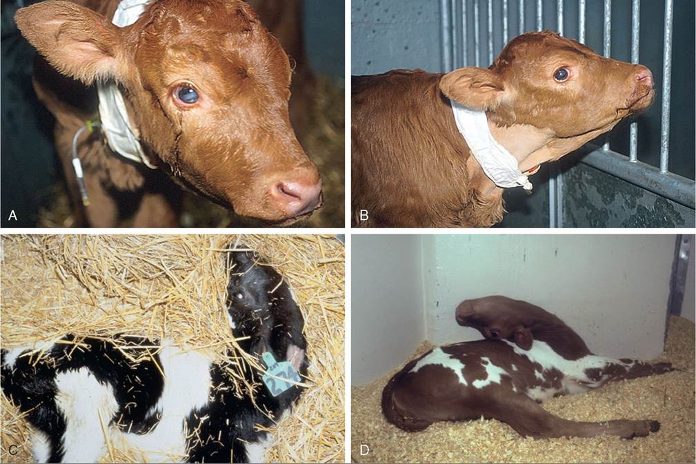
FIG. 20.1 Calves exhibiting clinical signs of meningitis secondary to septicemia. A, The calf is depressed and maintains an extended neck when manipulated. B, The presence of fibrin and white blood cells in the anterior chamber of the eye (hypopyon) is suspected. C and D, Calves with more advanced meningitis exhibiting profound opisthotonus. Images reprinted with permission from Dore V, Smith G. Cerebral disorders of calves. veterinary clinics of north america-food. Animal Practice 2017;33:27.)
■ TABLE 20.2
Cerebrospinal Fluid (CSF) Findings in Calves With Meningitis
| Normal | Green et al.29 | Scott et al. a | |
| Specific gravity | toxicity. In contrast, aminoglycosides diffuse relatively easily across the blood-brain barrier but likely represent a poor choice for meningitis, since standard doses are not high enough to achieve therapeutic concentrations in the CSF and higher-than-standard doses carry a high risk for nephrotoxicity.28 Although broad-spectrum antimicrobials are indicated, E. coli is the most common isolate from meningitis cases and so should be the primary target of initial therapy. The choice of antimicrobial for treating meningitis will depend largely on the veterinary drug laws in the country where the calf is being treated. For example, common recommendations for meningitis treatment in calves have included ceftiofur (5 to 10 mg/kg q8h IM or IV) or enrofloxacin (5 mg/kg q12h IV).20 Either of these would likely represent a reasonable option, but both are prohibited in the United States. Labeled doses of ceftiofur (2.2 mg/kg IM) could be used, or possibly sodium ampicillin (10-20 mg/kg q8h IV) could be tried. Another potential option would be potentiated sulfonamides such as trimethoprim-sulfamethoxazole dosed at 5 mg/kg (trimethoprim component) q8-12h IV. Potentiated sulfonamides could also be given concurrently with sodium ampicillin or ceftiofur to try to broaden the spectrum of activity. Intrathecal therapy (administering antibiotics directly into the CSF) has not been shown to improve outcome in human medicine and is likely too complicated for use in farm animals.31 In a CSF pharmacokinetic study of florfenicol in calves, the maximum concentration of florfenicol attained in CSF was 4.67 ± 1.51 pg/mk following a single intravenous dose of 20 mg/kg.26 The levels remained above the minimum inhibitory concentration (MIC) for Histopbilussomni over a 20-hour period. This concentration is below the MIC90 for E. coli. Bactericidal antibiotics are proposed to be more effective for treatment of meningitis in humans, and it is recommended that the concentration of antibiotic in the CSF should be maintained at 10 times the MIC of the target pathogen.30 Unfortunately, owing to the lack of CSF pharmacokinetic data in cattle, antimicrobial treatment of meningitis is an inexact science. Antiinflammatory therapy and nursing care should be instituted as described previously for neonatal septicemia. Convulsions in calves can be treated with diazepam (0.1 to 0.2 mg/kg IV). This can be repeated every 30 minutes until convulsions are controlled. Metabolic Acidosis Profound weakness associated with metabolic acidosis or “strong ion” acidosis is commonly observed in calves with diarrhea and sporadically in kids (“floppy kid syndrome”) and calves without other clinical signs of disease.32,33 This acidosis is often associated with an increased concentration of D-lactate (the anion of D-lactic acid) resulting from bacterial fermentation of carbohydrates in the GI tract of milk-fed calves. Clinical signs of impaired CNS function, including ataxia and coma, in sick calves have been attributed at least partially to the increased D-lactate concentrations.34,35Assessment of weak neonatal ruminants for the presence of metabolic acidosis is a critical part of the physical examination. Correction of the acidosis by intravenous administration of bicarbonate produces a rapid recovery. An improvement in mentation and strength should be observed within 12 hours; persistent depression is likely to reflect incomplete correction of acidosis, sepsis, hypoglycemia, hypernatremia, or hyponatremia. Hypoglycemia Hypoglycemia is a common sequela to withdrawal of milk for more than 48 hours, especially in cold weather. Affected calves are weak or recumbent but appear to be normally hydrated or minimally dehydrated.36 They are often emaciated and can occasionally have neurologic signs, including facial twitches, convulsions, opisthotonos, and coma. They will respond to a bolus of 50% dextrose or an infusion of 5% glucose, but often this response is temporary, especially in calves with severe malabsorptive disease. It is important to rapidly restore adequate energy intake to ensure resolution of these cases. Starvation and hypothermia resulting from mismothering are common causes of weakness in neonatal lambs. Similarly, weakness, poor body condition, and increased susceptibility to infectious diseases are observed with protein-calorie malnutrition induced by feeding poor-quality or incorrectly mixed milk replacers.37 Hypoglycemia is also frequently seen in calves with neonatal septicemia, as discussed previously. In a recent study that reported serum glucose concentrations in more than 10,000 calves 3 weeks of age and younger, the presence of severe hypoglycemia (glucose ≤36 mg/dL or 2 mmol/L) was associated with a poor survival rate, as only 20.6% of these calves survived compared to 74.0% of calves with normal glucose levels.38 Hyponatremia Hyponatremia occurs when loss of isotonic fluid through the GI tract is replaced by free water or hypotonic solutions. The latter often occurs when too much water is added when making up an oral electrolyte solution. Hyponatremia may also occur when isotonic oral electrolyte solutions are administered to calves with compromised sodium absorption capacity. This may be a result of severe pathologic changes or an inadequate level of agents that facilitate sodium cotransport within the oral electrolyte solution. Hyponatremia results in a fluid shift from the extracellular space to the intracellular compartment along the osmotic gradient, and the resultant swelling of the cells can result in neurologic disturbances, depression, disorientation, and even convulsions.39 Hyponatremia should be considered in calves with serum sodium less than 132 mmol/L; calves with serum sodium less than 120 mmol/L have severe hyponatremia. The goal of therapy is to restore serum sodium levels to greater than 125 mmol/L over the first 6 hours and then to restore to normal levels over 24 hours.39 In hypovolemic calves the initial treatment should be achieved using normal saline, and in normovolemic calves hypertonic saline should be used for the initial treatment, since the administration of large fluid volumes will exacerbate cerebral edema. If the calves are also suspected to be acidotic, this should be corrected with sodium bicarbonate solutions of appropriate tonicity. The amount of sodium required in the first 6 hours to raise the sodium level to 125 mmol/L can be calculated as follows.39 mEq Sodium = (125 - Measured mEq serum sodium) ? (0.6 ? Body weight in kg) Calves should then be maintained on a sodium-containing isotonic fluid, such as normal saline or lactated Ringer, and treated with oral electrolyte solution as appropriate. The sodium level should be monitored frequently in the first 24 hours because of unknown losses through the GI tract as well as unknown kidney function in a severely dehydrated patient. Hypernatremia Hypernatremia (also known as sodium or salt toxicity) is a relatively common problem encountered in young calves. These cases are generally a result of the calf ingesting excessive concentrations of salt without an adequate balance of water to balance the sodium load. The most common clinical scenarios involve calves ingesting milk replacers high in sodium without access to fresh water40,41 or calves that are being treated for diarrhea and receive oral electrolyte solutions with high sodium concentrations.42,43 Recently, cases of hypernatremia in calves have been observed associated with the use of high-sodium water on farms. Affected calves are typically younger than 3 weeks of age and often (but not always) exhibit neurologic signs. The volume of the extracellular fluid (ECF) is determined by the total body sodium content, whereas the osmolality and sodium concentration of the ECF are determined by sodium and water balance. Multiple control mechanisms work in the body to detect and respond to changes in the osmolality and volume of the ECF. These homeostatic mechanisms primarily control retention and excretion of sodium. Examples of regulatory control include the thirst response (water ingestion) and excretion or retention of sodium by the kidneys. Although it sounds simple, these are under complex control by multiple hormonal pathways, including antidiuretic hormone (ADH), atrial natriuretic peptide (ANP), the renin-angiotensin- aldosterone system (RAAS), and the autonomic nervous system. A complete discussion of the physiology of sodium and water homeostasis is beyond the scope of this chapter, but readers are referred to other recent review articles for more detailed information.44,45 When calves take in too much sodium, this results in expansion of the ECF at the expense of intracellular fluid (ICF) in order to return the osmolality of the blood to normal (relative dehydration). In cases where there is no water intake, fluid loss is associated with reduction in total body water content (absolute dehydration). In cases of hypernatremia, the brain has to adapt to the hyperosmolar state, which typically involves shrinkage of neurons as water follows concentration gradients and moves into the hyperosmolar CSF and plasma. This results in dehydration of neurons and brain shrinkage. As the brain moves away from the calvaria, disruption of the normal blood supply can occur.44 Neurologic signs often worsen during treatment of calves with hypernatremia, since rapidly lowering the sodium concentration with free water will allow water to flow into neurons, causing the already shrunken cells to swell, resulting in cerebral edema. This edema can lead to seizures and in many cases death.28 Hypernatremia is defined as a serum or plasma sodium concentration of 160 mEq/L or greater, and neurologic signs of salt toxicity can be seen. Serum sodium concentrations greater than 180 mEq/L generally indicate a guarded to poor prognosis. Sodium concentrations in the CSF of cattle normally range from 130 to 142 mEq/L but are typically above 160 mEq/L in animals with hypernatremia.46 Ocular fluid represents a good sample to submit to the diagnostic laboratory in dead calves, since it does not change appreciably with advancing postmortem autolysis and provides a stable representation of sodium levels at the time of death. Ocular fluid sodium concentration is reported to be about 95% of the serum sodium value, and levels are significantly elevated in cattle with hypernatremia.46 In cattle that are dead, the entire brain or cerebral cortex should be submitted to a diagnostic laboratory for sodium analysis and histopathology. Typically no gross lesions of the brain are apparent at necropsy in cases of hypernatremia, and histopathology is nonspecific with cerebral edema present. Therefore submitting fresh brain and/or ocular fluid for sodium analysis in addition to requesting histopathology is critical. Sodium concentrations in brain tissue are generally greater than 2000 μgλg (ppm) in calves with hypernatremia. Early clinical signs of hypernatremia include lethargy and depression, which, without blood work, cannot be differentiated from many other possible diseases such as acidemia, dehydration, hypoglycemia, or hypothermia. More advanced clinical signs of hypernatremia include twitching of facial muscles, muscle rigidity, tremors, and myoclonus.28,44,47 Calves will demonstrate seizure and/or coma activity near death. Prognosis in these calves is often guarded to poor even with aggressive treatment. Treatment of calves with chronic hypernatremia is often very difficult and should be reserved primarily for valuable animals. Classic recommendations call for sodium concentrations to be lowered very slowly, since replacing free water too rapidly could cause neurons to swell, cerebral edema, and a worsening of neurologic signs. The total body free water deficit can be calculated using the following formula: 0.6 ? Body weight (kg) ? ([Current serum sodium concentrations/Normal serum sodium concentration] -1) However, if this fluid is given rapidly, neurologic complications will often arise. In general the recommendation is to formulate fluids that have a sodium concentration approximately equal to that of the calf.28,44,47 This can be done by adding small volumes of hypertonic saline (1.2 mEq/mL) or 23.4% sodium chloride (4 mEq/mL) to whatever fluid type you have chosen to give the calf. For example, if the calf has an acidemia, additional sodium can be added to isotonic sodium bicarbonate (sodium concentration of 156 mEq/L). However, if the calf has a relatively normal pH, additional sodium could be added to 0.9% saline (sodium concentration of 154 mEq/L). The idea is to decrease the sodium level of the calf very slowly over several days to avoid cerebral edema. A case example below will illustrate the calculation. A 45-kg Hereford calf had clinical signs of hypernatremia. The calf had a serum sodium concentration of 187 mEq/L. To calculate the free water deficit in this calf we would multiple 0.6 ? 45 ? ([187 - 145) - 1)] to get a free water deficit of 7.82 L. In this example we used 145 to represent the “normal” serum sodium concentration for a calf. If we wanted to give 0.9% saline to this calf (154 mEq of sodium per liter), we would need to add 33 mEq of additional sodium to reach the calf's level of 187. The simplest way to do this would be to add a small volume of hypertonic saline (33 mEq/1.2 mEq/ mL = 27 mL of hypertonic saline added per liter of fluids). You could also add hypertonic saline to isotonic sodium bicarbonate if the calf had an acidemia. Since isotonic sodium bicarbonate has a sodium concentration of 156 mEq/L, you would need to add 31 mEq of sodium. Be careful about adding large volumes of dextrose to these solutions, as that will lower the sodium concentration. Although not always successful, a report of treating several hypernatremic calves with 5% dextrose either alone or in combination with sodium bicarbonate has been published.42 In this study calves were given 2 to 4 L of 5% dextrose at a slow drip (about 500 mL/h) and had their serum sodium concentrations decreased much faster than is usually recommended. However, the four calves in the case report were treated successfully. Although this approach may not always work and could easily result in cerebral edema, it may be practical for times when the clinician does not have the ability to formulate fluids or monitor serum sodium concentrations frequently. In calves with chronic hypernatremia where “idiogenic osmoles” have formed, it is likely critical to lower sodium levels slowly, whereas the clinician may be able to lower sodium more rapidly in cases of acute hypernatremia. If the calf's neurologic signs get significantly worse after fluid therapy has started, cerebral edema should be suspected. Treating cerebral edema is difficult but imperative to prevent further brain damage and/or death. Corticosteroids are the easiest choice normally; however, they are only marginally effective in treating moderate to severe cases of cerebral edema. Mannitol (25%) at 1 g/kg given IV over 30 minutes or an oral solution of glycerin diluted 1 : 1 with water has also been recommended for severe cases of cerebral edema but can be more difficult to obtain.28 Hypernatremia is largely avoidable by proper management. Since hypernatremia occurs largely under one of two condi- tions—(1) calf ingests too much sodium without an adequate volume of water in a short period of time, or (2) sustained water deprivation—the key becomes preventing both of these conditions from occurring. Whole milk is typically fairly low in sodium concentration, so typically calves fed whole milk have little problem with sodium toxicity unless given a large amount of oral electrolytes. Some milk replacer products can have significantly higher sodium concentrations as compared to whole milk (up to 85 to 100 mmol/L). Water quality is another factor that can have a profound effect on the amount of sodium calves ingest. Water that has been through a softening process to remove minerals such as calcium and magnesium can often have very high sodium concentrations and therefore should not be used to dilute commercial milk replacers unless a water analysis has been performed and sodium levels are less than 100 ppm. In one case report of salt poisoning in calves, milk replacer samples diluted with farm water had sodium concentrations ranging from 171 to 185 mmol/L.40 When cases of hypernatremia are diagnosed on a farm, veterinarians should not forget to consider the drinking water source as a possible source of excess sodium. In addition to watching how much sodium we feed calves in their normal milk diet, attention must also be given to the feeding of oral electrolyte solutions in calves with diarrhea. When homemade oral electrolyte products are used or when labeled mixing directions are not followed correctly, sometimes the sodium concentration fed to calves can be extremely high. In some cases oral electrolyte products are purchased in bulk containers, and more powder is added to 2 L of water than indicated in the directions. Multiple case reports describing hypernatremia in calves have involved the feeding of unknown amounts of oral electrolyte solution to calves multiple times per day.42,43 This situation is exacerbated when calves do not have access to fresh water or when the fresh water also contains high sodium concentrations. In one case report, hypernatremia was diagnosed associated with neurologic signs and deaths in a group of 6- to 10-month-old Holstein calves that had access to a salt block without free-choice water available.48 So ultimately, paying attention to the calf feeding program and the diarrhea treatment (oral electrolyte) feeding protocol for the farm is critical, along with ensuring that calves always have a supply of fresh, good-quality water. Neuromuscular and Musculoskeletal Disease Primary neuromuscular or musculoskeletal disease should be considered when weakness is not associated with depressed mentation. Weakness associated with micronutrient deficiencies results from myodegeneration (white muscle disease, selenium, and vitamin E) or demyelination (copper, enzootic ataxia). If weakness is detected in one or more limbs immediately after birth, peripheral nerve and muscle damage associated with birth trauma should be ruled out (see Box 20.1). Femoral nerve paralysis may be observed in calves following a “hip lock” dystocia.49 A condition resembling congenital myasthenia gravis has also been described in Brahman calves.50 Nutritional myodegeneration associated with selenium or vitamin E deficiencies may produce localized (dysphagia) or generalized paresis. Neonatal small ruminants appear to be particularly susceptible. Affected lambs may be unable to rise; others can stand but may be unable to nurse because they are unable to raise their heads. Diagnosis is based on clinical signs, increased serum creatine kinase concentration, and reduced whole blood glutathione peroxidase and/or selenium concentrations (see Chapter 42). Vitamin E deficiency is observed when pregnant ewes are fed stored forage low in vitamin E; the clinical signs in affected lambs are identical to selenium deficiency, but selenium status is adequate. Since vitamin E is labile, serum should be harvested quickly after blood collection, frozen, wrapped in aluminum foil, and sent via express mail on ice. Paraplegia and tetraplegia are commonly associated with spinal cord compression. Compression of the spinal cord in neonates most commonly results from vertebral body malformations, osteomyelitis, or fractures. Generally vertebral body malformations occur sporadically; genetic, nutritional, and environmental factors have been implicated.51,52 In older calves, underlying metabolic bone disease (copper, vitamin D, or phosphorous deficiency) may increase the propensity for fractures to occur. Osteomyelitis and vertebral body abscess may be sequelae to bacteremia following neonatal septicemia or pneumonia.53 The frequent isolation of Trueperella pyogenes from vertebral body abscesses in ruminants suggests that chronic respiratory infection is more frequently the source in these species.54,55 Vertebral body abscesses in lambs are occasionally a sequela to infected docking wounds. Leukocytosis and hyperfibrinogenemia are commonly observed in neonates with vertebral body abscesses. In most instances vertebral abscesses do not infiltrate the pachymeninges, so the CSF either is normal or has a mild elevation of protein and/or a mild pleocytosis.53,54 Differential diagnoses for paresis in goat kids include caprine arthritis encephalitis virus (CAEV) and enzootic ataxia. Enzootic ataxia is also common in lambs. Progressive ataxia and paresis or paralysis is a feature of both diseases. There are two forms of enzootic ataxia (swayback): the neonatal and the delayed types. In the neonatal condition, animals are affected at birth; in the delayed type, signs of incoordination appear at 14 to 30 days of age.56 Most affected kids are afebrile, bright, and alert, and they will continue to eat if it is physically possible. Enzootic ataxia is associated with low liver copper content and occasionally low serum copper concentration. It has been proposed that reduction in the activity of the copper-dependent enzyme cytochrome oxidase impairs phospholipid synthesis and subsequently myelin production. Microcytic anemia and increased fragility of bones may be observed in more chronic cases.57 The copper, molybdenum, and sulfur content of the maternal diet should be evaluated and adjustments made for copper deficiency or molybdenum or sulfur excess. Goat kids with the neurologic form of CAEV will have mild to moderate fevers and evidence of cerebral involvement. Cerebral signs commonly identified include depression, head tilt, torticollis, and circling. Evidence for CAEV includes CSF pleocytosis and increased CSF protein and a positive CAEV (agar gel immunodiffusion, or AGID) or enzyme-linked immunosorbent assay (ELISA) test.58 Both the neurologic form of CAEV and enzootic ataxia carry a poor prognosis. A complete neurologic examination is an important component of the work-up of the weak neonate. In particular, it should be noted if the weakness is accompanied by signs of depression and diffuse cerebral disease. Strength is preserved if ataxia is caused by cerebellar disease. Limb reflexes should be tested to establish whether components of the spinal reflex pathways are involved in the disease process (sensory nerve, lower motor neuron, neuromuscular junction, muscle). Animals with other types of spinal cord disease (e.g., trauma, vertebral malformations, enzootic ataxia) may also show weakness and ataxia yet appear clinically to have normal cerebral function. Virtually any severe systemic disease such as generalized infection can cause both profound depression and weakness in a neonate without the presence of actual brain pathology. Intermittent signs of severe weakness and depression may be caused by the narcolepsy-cataplexy syndrome.
More on the topic Weakness and/or Depressed Mentation:
-
Veterinarian -
|