Accessory Digestive Organs
in addition to the numerous small glands located in the walls of the stomach and intestine, accessory glands include the salivary glands, the pancreas, and the liver.
Salivary Glands
The salivary glands of domestic farm animals comprise three pairs of well-defined glands as well as scattered lobules of salivary tissue (minor salivary glands).
The chief salivary glands are the parotid, mandibular, and sublingual (Fig. 20-15). The minor salivary glands include labial, buccal, lingual, and palatine glands.The parotid salivary gland is located ventral to the ear in relation to the caudal border of the mandible. In most animals, the parotid salivary duct passes ventrad and craniad on the deep face of the caudal part of the mandible and crosses the cheek superficially just cranial to the masseter muscle. The duct then passes dorsad to penetrate the mucous membrane of the cheek near the third or fourth maxillary cheek tooth.
The mandibular salivary gland is usually located ventral to the parotid gland, just caudal to the mandible. The mandibular salivary duct passes forward along the medial side of the mandible to open ventral to the tongue on the sublingual caruncle, a small hillock of mucous membrane located on the floor of the mouth.
The sublingual salivary gland is located deep to the mucous membrane along the ventral side of the lateral surface of the tongue near the floor of the mouth. Numerous ducts pass directly from the gland to open into the floor of the mouth just ventrolateral to the tongue. With the exception of the horse, the sublingual salivary gland also has a monostomatic portion that empties at the sublingual caruncle on the floor of the mouth by way of a single sublingual duct that runs parallel to the mandibular duct.
The salivary glands are classified as serous, mucous, or mixed. serous glands secrete a watery clear fluid, as compared with mucous glands, which secrete mucus, a viscous material that acts as a protective covering for the surface of mucous membranes.
A mixed gland produces both mucous and serous fluids. The parotid salivary gland secretes primarily serous saliva; mandibular and sublingual glands are classified as mixed glands in domestic farm animals. Most of the minor salivary glands have a mucous secretion.Pancreas
The pancreas is a compound gland that has both endocrine and exocrine portions. The exocrine portion of the pancreas produces sodium bicarbonate and digestive enzymes, which pass through the pancreatic ducts to empty into the duodenum close to the opening of the bile duct.
The endocrine portion of the pancreas consists of isolated groups of pale-staining cells scattered throughout the gland. These areas are called the pancreatic islets (formerly islets of Langerhans). They produce the hormones that pass directly into the bloodstream (see Chapter 12), most notably glucagon and insulin, which are the primary regulators of blood sugar levels.
Grossly, the pancreas is an irregularly lobu- lated organ that lies adjacent to the proximal duodenum and frequently abuts the stomach, the caudal vena cava, and caudal part of the liver as well. The pancreas has the appearance of aggregated nodules loosely connected to form an elongated gland lying parallel to the duodenum. The developing pancreas arises as two diverticula of the embryonic duodenum and therefore always begins as a bilobed organ connected to the lumen of the gut by two ducts. During organogenesis in many species, the duct systems of the two lobes intermingle and one of the two original connections to the gut lumen is lost; thus, a single duct is the normal condition of the adult in these species. The disposition of ducts of the pancreas is shown in Table 20-3.
in those species that possess it, the pancreatic duct opens onto a small elevation within the duodenum in common with the bile duct from the liver. This is the major duodenal papilla. A short distance away, a smaller minor duodenal papilla marks the location of the accessory pancreatic duct.
As small ruminants lack the accessory pancreatic duct, they also lack the minor duodenal papilla.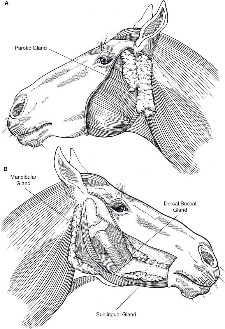
Figure 20-15. Major salivary glands. A) Superficial view. B) Deep view, with mandible cut away.
Table 20-3. Species Variations in Pancreatic Ducts
| Species | Pancreatic Duct | Accessory Pancreatic Duct |
| Horse | + | + |
| Ox | Usually absent | + |
| Pig | - | + |
| Small ruminants | + | — |
The first branches of these ducts within the pancreas are interlobular ducts, so called because they run between lobules of the pancreas. Interlobular ducts branch into intralobular ducts that enter individual lobules and give rise to intercalated ducts, which enter the acini (sometimes called alveoli).
Liver
The liver is the largest gland in the body, constituting 1-2% of total adult body weight. It varies somewhat in number of lobes and precise intra-abdominal location from one species to another. However, the liver is always located immediately caudal to the diaphragm (in contact with it) and tends to be located on the right side, particularly in ruminants, in whom the large ruminoreticulum pushes everything else to the right.
Individual liver lobules of the pig are encircled by rather heavy connective tissue septa, which give a lobulated, “cobblestone” appearance to the surface of the porcine liver. This appearance is less distinctive in other domestic species. Liver tissue is usually a reddish brown, although accumulation of fat (whether due to a high-fat diet or pathology) can give it a pronounced yellow tinge.
The liver receives two blood supplies. To provide oxygen and nutrients, arterial blood from the hepatic artery, a branch of the celiac artery, enters the side of the liver adjacent to the viscera, called the porta (the Latin word for gate). This is the nutrient blood supply. The porta also receives the large portal vein, which carries blood to the liver from the stomach, spleen, pancreas, and intestines. The liver per-
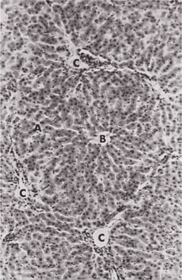
Figure 20-16. Microscopic anatomy of the bovine liver. A, Hepatocytes of liver lobule; B, central vein; C, portal veins. (Reprinted with permission of Wiley- Blackwell from Dellmann H.D. Textbook of Veterinary Histology. 4th ed. Philadelphia: Lea & Febiger, 1993.)
forms metabolic and immunologic functions on this blood returning from the gastrointestinal tract, and so the blood of the portal vein constitutes the functional blood supply. Portal blood is detoxified and modified within the sinusoids (capillaries) of the liver and then leaves the liver by way of the short hepatic veins that empty into the caudal vena cava.
All domestic animals except the horse have a gallbladder for storage of bile. The liver’s digestive secretion, bile, leaves the liver through hepatic ducts, which join the cystic duct from the gallbladder to form the common bile duct, which then passes to proximal duodenum into the lumen to which it opens in common with the pancreatic duct on the major duodenal papilla (see above).
Microscopically, the morphologic unit of the liver is the hepatic lobule, a polygonal cylinder of liver cells (the hepatocytes) in the center of which is a central vein (Fig. 20-16). At the angles on the periphery, where adjacent hepatic lobules meet, are the portal triads, consisting of branches of the hepatic artery and portal vein (interlobular vessels), an interlobular bile duct, and lymphatics. These communicate with the spaces between sheets, or laminae, of hepatocytes in the hepatic lobule; the spaces are the hepatic sinusoids, and they are characterized by the lack of a basal lamina and numerous fenestrations in the endothelium, allowing free egress of blood constituents that then bathe the hepatocytes. Blood (both arterial and portal) flows from the portal canal through the sinusoids and is gathered by the central vein, the smallest tributary of the hepatic veins. in and around the sinusoids are fixed macrophages, which in this location are called Kupffer cells.
Between adjacent rows of liver cells is a tiny bile canaliculus, which is little more than a tube formed by grooves in the surfaces of the apposed liver cells. Bile produced by the hepatocytes is carried toward the periphery of the hepatic lobule by the bile canaliculi to the interlobular bile ducts located at the portal canal (notice that this net flow is opposite the direction of blood flow).