For normal metabolism, cells of an animal’s body need the three major classes of nutrients (carbohydrates, proteins, and lipids) delivered to them via the blood in their simplest forms (monosaccharides, amino acids, and fatty acids).
Animals consume foodstuffs that contain these nutrients in more complex chemical and physical forms. It is the function of the gastrointestinal tract to reduce the consumed foodstuffs to simpler molecules and to transfer them to the blood so that they can be delivered to the cells for metabolism.
The processes of physical and chemical breakdown of foodstuffs are termed mechanical and chemical digestion, respectively. In addition to monosaccharides, amino acids, and fatty acids, the gastrointestinal tract must absorb other essential minor nutrients (e.g., salts, vitamins), so that they are available to the cells of the body.The gastrointestinal tract is essentially a long, smooth muscle tube extending from mouth to anus. The tube has two distinct layers of smooth muscle in its wall (circular and longitudinal layers) and is lined with epithelia that function as selective barriers between the lumen and the body fluids. The anatomic and functional characteristics of the mucosa and its epithelia vary greatly among segments of the intestine (Fig. 21-1). Indigestible substances or items (such as a coin) can pass through the tract without being altered and without affecting the
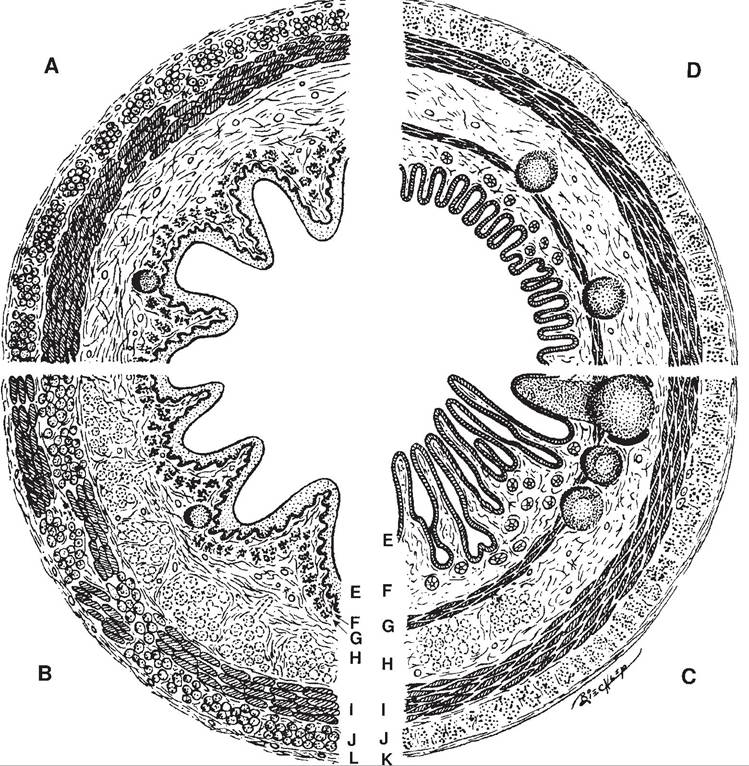
Figure 21-1. Cross-sections through various segments of the digestive tract. A and B, Esophagus with stratified squamous epithelium. C, Small intestine with columnar epithelium and submucosal glands and aggregated lymphatic nodules in some segments. D, Large intestine. E, tunica mucosa: epithelium; F, lamina propria; G, lamina muscularis; H, tela submucosa; i, tunica muscularis: circular layer; J, longitudinal layer; K, tunica serosa; L, tunica adventitia. (Reprinted with permission of Wiley-Blackwell from Dellmann H.D. and Eurell J. Textbook of Veterinary Histology.
5th ed. Philadelphia: Lippincott Williams & Wilkins, 1998.)animal if they are not large enough to impair movement of the other contents.
The smooth muscle in the wall of the gastrointestinal tract provides the force to move digesta through the tract; gastrointestinal motility is the general term used to describe the activity of this smooth muscle. Gastrointestinal motility is primarily regulated by three mechanisms: (1) autonomic nervous system, (2) gastrointestinal hormones, and (3) enteric nervous system.
Gastrointestinal hormones are released from endocrine cells in the epithelial lining of the gastrointestinal tract (enteroendocrine cells) and may stimulate or inhibit gastrointestinal smooth muscle. The release of these hormones is usually in response to digesta in the lumen of the tract. Thus, these hormones are a means of local regulation that is coordinated with the ingestion and digestion of food.
The enteric nervous system consists of neural plexuses between layers of smooth muscle in the wall of the tract (Fig. 21-1). These plexuses contain complete neurons (dendrites, cell bodies, and axons) that can form complete neural and reflex circuits in the wall of the tract so that neural regulation can be independent of external innervation. The presence of food and distension of gastrointestinal tract segments act as stimuli to initiate activity of the enteric nervous system. The three regulatory mechanisms (autonomic nervous system, gastrointestinal hormones, and enteric nervous system) also regulate secretions from glands in the wall of the gastrointestinal tract (Fig. 21-1) and the intestinal accessory organs (salivary glands, liver, and pancreas).
All three of these mechanisms may regulate a given intestinal segment or accessory organ, but the relative importance of each varies among the segments of the gastrointestinal tract and the accessory organs. For example, salivary secretion is almost entirely regulated by the autonomic nervous system, whereas gastrointestinal hormones are primary in the initiation of bile secretion.