Although the appropriate regulation of “visceral” activities clearly presumes the existence of receptors in the
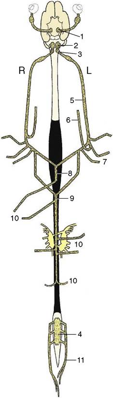
Figure 8-73 Origin and distribution of the parasympathetic nervous system. Ventral view, schematic.
1, Parasympathetic oculomotor nucleus; 2, rostral and middle parasympathetic nuclei of the medulla oblongata; 3, dorsal vagal nucleus; 4, sacral outflow; 5, vagus nerve; 6, recurrent laryngeal nerve; 7, parasympathetic fibers to heart and lungs; 8, ventral vagal trunk; 9, dorsal vagal trunk; 10, parasympathetic fibers to the abdominal organs; 11, pelvic nerves.viscera and vessels, the autonomic nervous system was originally defined as wholly efferent. This offers a certain convenience because visceral afferent pathways are in general indistinguishable in structure and arrangement from their somatic counterparts. The visceral efferent pathways, on the other hand, are clearly distinguished, particularly by the location of the last neuron in the chain within a peripheral ganglion and by the restriction of the neurons that drive these ganglion cells to specific nuclei of the brainstem and particular regions of the cord (Figures 8-73 and 8-74). The peripheral
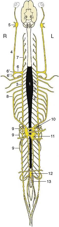
Figure 8-74 Origin and distribution of the sympathetic nervous system. Ventral view, schematic. The parasympathetic nuclei in brain and spinal cord are indicated in gray. 1, Sympathetic outflow from T1 to L3; 2, communicating branches; 3,4, sympathetic trunk; 5, cranial cervical ganglion; 6, cervicothoracic ganglion; 6', middle cervical ganglion; 6", ansa subclavia; 7, vertebral n.; 8, greater splanchnic n.; 9, lesser splanchnic nn.; 10, celiac ganglion; 11, cranial mesenteric ganglion; 12, caudal mesenteric ganglion; 13, hypogastric n.
efferent pathway thus consists of a preganglionic (myelinated, and therefore white) fiber and a postganglionic (little myelinated, and therefore gray) fiber.
Moreover, certain anatomical, physiological, and pharmacological features distinguish two contrasting— sympathetic and parasympathetic—efferent systems, whereas no similar distinction is possible for the visceral afferent fibers presumed to be included in all cranial and spinal nerves (if only because of the ubiquitous distribution of blood vessels). It has been shown (p. 304) that cerebrospinal (somatic) and autonomic (visceral) mechanisms cannot be entirely separated because the cerebral cortex directs both types.Some contrasting physiological actions of the two systems are summarized later (p. 331), but it may be said now that they partly rest on the use of norepinephrine as the mediating substance at the last synapse of the sympathetic pathway, while acetylcholine is used at the corresponding parasympathetic synapse. Epinephrine is produced by the adrenal medulla, and when generally diffused by the bloodstream, it evokes a mass sympathetic response. Acetylcholine is liberated and destroyed locally. The activities of the parasympathetic system therefore tend to be more specific and discrete than those of the sympathetic system. The narrower localization of parasympathetic responses is further assisted by the location of parasympathetic ganglia close by or even within the target organ, whereas the sympathetic ganglia are closer to the central nervous system and the postganglionic fibers radiate more widely.