Alveolar Macrophages Scavenge Particles Deposited on the Alveolar Surface
Macrophages, which constitute the majority of cells in the alveolar lining fluids, are the principal resident phagocytes in the normal lung. Macrophages originate in bone marrow as monocytes and differentiate during their passage from the blood into the alveolus, where their turnover time is in days.
Surfactant proteins, complement, opsonins, and lysozymes in respiratory tract secretions assist macrophages in the killing and removal of viable particulates, such as bacteria. Once phagocytized, particles are destroyed by the macrophage or transported out of the lung. Some macrophages enter the mucociliary system directly from the alveolus; others traverse the alveolar wall and enter the lymphoid tissues associated with the airways. In the lymphoid tissue, macrophages are antigen-presenting cells (APCs) and thus play a critical role in orchestrating the lungs immune responses.Macrophages have adapted to the high oxygen levels of the alveolus, and their role as phagocytes is depressed by hypoxia. Macrophage function is also suppressed by endogenous glucocorticoids that are released from the adrenal glands at times of stress and by synthetic corticosteroids that are used to relieve inflammation (e.g., in arthritis). Stress-induced suppression of macrophage function contributes to respiratory disease in animals transported for long distances. In addition, excessive administration of synthetic corticosteroids can make animals more susceptible to bacterial infections of the lung. Viral infections also suppress macrophage function; this occurs approximately 7 days after virus inoculation (Figure 50-3). This contributes to the secondary bacterial infections that usually follow viral respiratory disease.
Alveolar macrophages are a first line of defense. When large numbers of particles are inhaled, other phagocytes from the blood, particularly polymorphonuclear leukocytes, especially
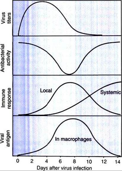
FIGURE 50-3 Effects of viral infection on antibacterial activity of alveolar macrophages.
Antibacterial activity is depressed 7 days after experimental viral infection. At this time, the viral antigen is located in the macrophages, which are damaged by the local immune response to the virus. (From Jakab GT: Viral-bacterial interactions in respiratory tract infections: a review of the mechanisms of virus-induced suppression of pulmonary anti-bacterial defenses. In Loan RW, editor: Bovine respiratory disease: a symposium, College Station, 1984, Texas A&M University Press.)neutrophils (PMNs), assist the macrophage. Toxic oxygen radicals and proteolytic enzymes are released by phagocytic cells to break down invading bacteria, but they may also damage the lung tissue in the process. Protease inhibitors (e.g., αl-antitrypsin) and antioxidants (e.g., glutathione peroxidase, ascorbic acid) protect the lung from its own defense mechanisms.