Anatomic language must be precise and unambiguous.
In an ideal world each term would have a single meaning, each structure a single name. Unhappily, there has long been an alarming superfluity of terms and much inconsistency in their use.
In the hope of reducing this confusion, an internationally agreed-on vocabulary—Nomina Anatomica Veterinaria (NAV)*-was introduced in 1968 and has since obtained wide acceptance. It was revised most recently in 2012, and we have tried to use it consistently throughout this work. Occasionally, we have included a second, older, and unofficial alternative when such a term appears to be so deeply rooted in clinical use that it is unlikely to be eliminated by edict. The terms of the NAV are in Latin, but it is permissible to translate them into vernacular equivalents. We have used translations that closely resemble the original Latin and give the Latin name only when the translation could be in doubt or there is no handy English equivalent. Because the names, whether in Latin or in English, are intended to be informative and an aid to comprehension, the reader should look terms up in a medical dictionary when their meaning is not self-evident.The terms that indicate position and direction must be mastered at once. These official terms are more precise than the common alternatives because they retain their relevance regardless of the actual posture of the subject. They are defined in the following list, and their use is illustrated in Fig. 1.1. We have not used them pedantically when there is no reasonable prospect of misunderstanding. When we use common terms (above, behind, and so forth), we always have in mind a standard anatomic position, which, for a quadruped, is that in which the animal stands square and alert. It differs from the human anatomic position. Medical anatomists make much use of the terms anterior and posterior and superior and inferior, all of which have very different connotations when applied to quadrupeds.
These terms are therefore best avoided, except for a few specific applications to the anatomy of the head.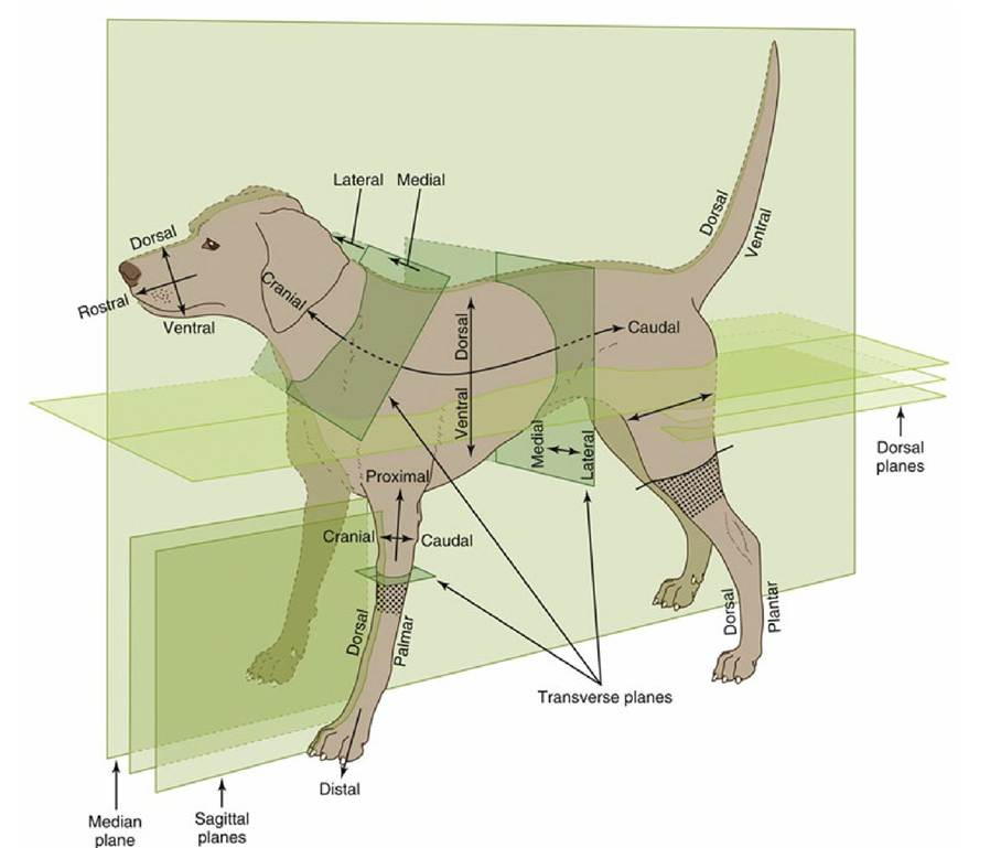
FIG. 1.1 Directional terms and planes of the animal body. The stippled areas represent the carpus and tarsus on forelimbs and hindlimbs, respectively.
The principal recommended terms of position and direction are arranged in pairs, and it should be emphasized that they refer to relative, not absolute, positions. Most of these adjectives, which are listed here, form corresponding adverbs with the addition of the suffix ly.
Dorsal structures (or positions) lie toward the back (dorsum) of the trunk or, by extension, toward the corresponding surface of the head or tail.
Ventral structures lie toward the belly (venter) or the corresponding surface of the head or tail. Cranial structures lie toward the head (cranium, literally skull), caudal ones toward the tail (cauda).
Within the head, structures toward the muzzle (rostrum) are said to be rostral; caudal remains appropriate.
Medial structures lie toward the median plane (medianus, in the middle) that divides the body into symmetrical right and left "halves."
Lateral structures lie toward the side (latus, flank) of the animal.
Different conventions apply within the limbs. Structures that lie toward the junction with the body are proximal (proximus, near), whereas those at a greater distance are distal (distantia, distance). Within the proximal part of the limb (which is defined for this purpose as extending to the proximal limit of the carpus [wrist] or tarsus [hock, ankle]), structures that lie toward the "front" are said to be cranial, those that lie toward the "rear" caudal. Within the remaining distal part of the limb, structures toward the "front" are dorsal (dorsum, back of the hand), and those toward the "rear" are palmar (palma, palm of the hand) in the forelimb or plantar (planta, sole of the foot) in the hindlimb. Additional terms may be applied to the anatomy of the digits. Axial structures lie close to the axis of a central digit, close to the axis of the limb if this passes between two digits; abaxial (ab, away from) positions are at a distance from the reference axis.
The terms external and internal, superficial, and deep (profundus) hardly require explanation or
definition.
Sometimes it is necessary to refer to a section through the body or a part of it (see Fig. 1.1). The median plane divides the body into symmetrical right and left halves. Any plane parallel to this is a sagittal plane, and those close to the median are sometimes termed paramedian planes. A dorsal plane sections the trunk or other part parallel to the dorsal surface. A transverse plane transects the trunk, head, limb, or other appendage perpendicular to its own long axis.