Anatomy of the Kidney
The kidneys are paired reddish-brown organs that filter plasma and plasma constituents from the blood and then selectively reabsorb water and useful constituents from the filtrate, ultimately excreting excesses and plasma waste products.
The kidneys of most animals are roughly bean-shaped, with the exceptions among domestic animals of the heart-shaped right equine kidney and the distinctively lobated kidneys of the ox (Figs. 23-1 and 23-2).The kidneys are in the dorsal part of the abdominal cavity on each side of the aorta and caudal vena cava, just ventral to the first few lumbar vertebrae. In most domestic animals, the right kidney is slightly more cranial than
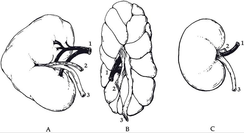
Figure 23-1. Right kidneys of the horse (A); the ox (B); and the sheep (C). 1, Renal artery; 2, renal vein; 3, ureter. (Reprinted with permission of Wiley-Blackwell from Reece W.O. Physiology of Domestic Animals. 2nd ed. Baltimore: Williams & Wilkins, 1997.)
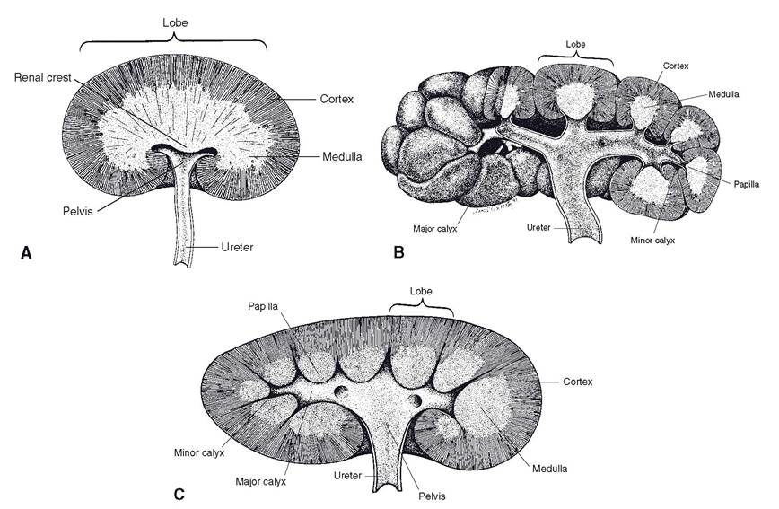
Figure 23-2. Internal anatomy of kidneys. A) The renal cortex of the equine, ovine, and caprine kidney lacks visible divisions into individual lobes. The renal papillae are fused in these species into a longitudinal renal crest. B) The bovine kidney is grossly divided into lobes, each of which communicates with a minor calyx. C) The porcine kidney lacks external divisions into lobes, but the renal papillae of the medulla distinguish each lobe internally. (Reprinted with permission of Wiley-Blackwell from Dellmann H.D. Textbook of Veterinary Histology. 4th ed. Philadelphia: Lea & Febiger, 1993.)
the left, with the cranial pole of the right kidney lying snugly in a complementary fossa of the liver. The left kidney tends to be more pendulous, and in ruminants, the forestomach may push the left kidney to the right as far as the median plane or beyond, particularly when the rumen is full.
The kidneys are described as being retroperitoneal in location, reflecting their position outside the peritoneal cavity where they are more closely attached to the abdominal wall by fascia, vessels, and peritoneum than are most other abdominal organs. A tough connective tissue capsule surrounds the entire kidney.The medial aspect of each kidney features a concavity, the hilus, where arteries and nerves enter the kidney, and the ureter, veins, and lymphatic vessels leave (Fig. 23-3). The wide origin of the ureter in the kidney is the renal pelvis. The renal pelvis receives urine from the collecting tubules of the kidney. The cavity in the kidney that contains the pelvis is the renal sinus. The bovine kidney does not have a renal pelvis, the ureter instead arising directly from the coalescence of individual calyces (discussed later).
The portion of the kidney immediately surrounding the renal pelvis is the renal medulla, which appears striated because of the radially arranged collecting tubules. In addition to collecting tubules, the medulla also contains some loops of Henle (descending and ascending loops). The medulla is surmounted peripherally by the renal cortex, in which reside the renal corpuscles, the histological units of filtration. The cortex has a granular appearance because of the large number of these renal corpuscles; also found in the cortex are proximal and distal convoluted tubules and other segments of loops of Henle (discussed later).
The medulla and cortex are arranged in units called lobes, cone-shaped aggregates of renal tissue. The medullary portion of each lobe constitutes a renal pyramid, whose apex, the renal papilla, is directed toward the renal pelvis (Fig. 23-3). In the bovine kidney, each pyramid is associated with one of the grossly obvious lobes of the bovine kidney. In the pig and small ruminants, the adjacent cortices of individual lobes are fused, so that the surface of the kidney appears smooth. The individual nature of the porcine lobes is revealed, however, through the persistence of discrete papillae projecting into the renal pelvis (Fig.
23-2). In the horse and small ruminants, the individual papillae, like the cortex, are fused. Consequently, they present as a single longitudinal ridge, the renal crest, projecting into the renal pelvis. Urine discharged from the collecting tubules of the renal crest is collected in the renal pelvis and from there is delivered to the ureter.In the kidney of the ox and pig, individual pyramids project into minor calyces, cuplike diverticula of the common collecting space within the renal hilus. These in turn empty into major calyces. These major calyces in the porcine kidney empty into the renal pelvis, but the bovine kidney has no pelvis, and so the major calyces in this species empty directly into the ureter (Fig. 23-2).
Blood and Nerve Supply
Because of its important role in adjusting the composition of extracellular fluid (including plasma), the blood supply to the kidney is much more extensive than the size of the organ would suggest. The two renal arteries may receive as much as one-fourth of the total cardiac output. Each renal artery enters the hilus of the kidney and divides into a number of relatively large branches, the interlobar arteries. These pass peripherally between pyramids almost to the cortex, where they bend abruptly and become arcuate arteries, which derive their name from the arched manner by which they pass along the junction between cortex and medulla (Fig. 23-3.)
Each arcuate artery gives off a number of interlobular arteries that extend into the cortex and in turn give rise to the afferent arterioles. Each afferent arteriole branches repeatedly to form a tufted capillary network called the glomerulus, which is associated with the renal corpuscle. The capillaries of the glomerulus coalesce into an efferent arteriole, which leaves each glomerulus (Fig. 23-4).
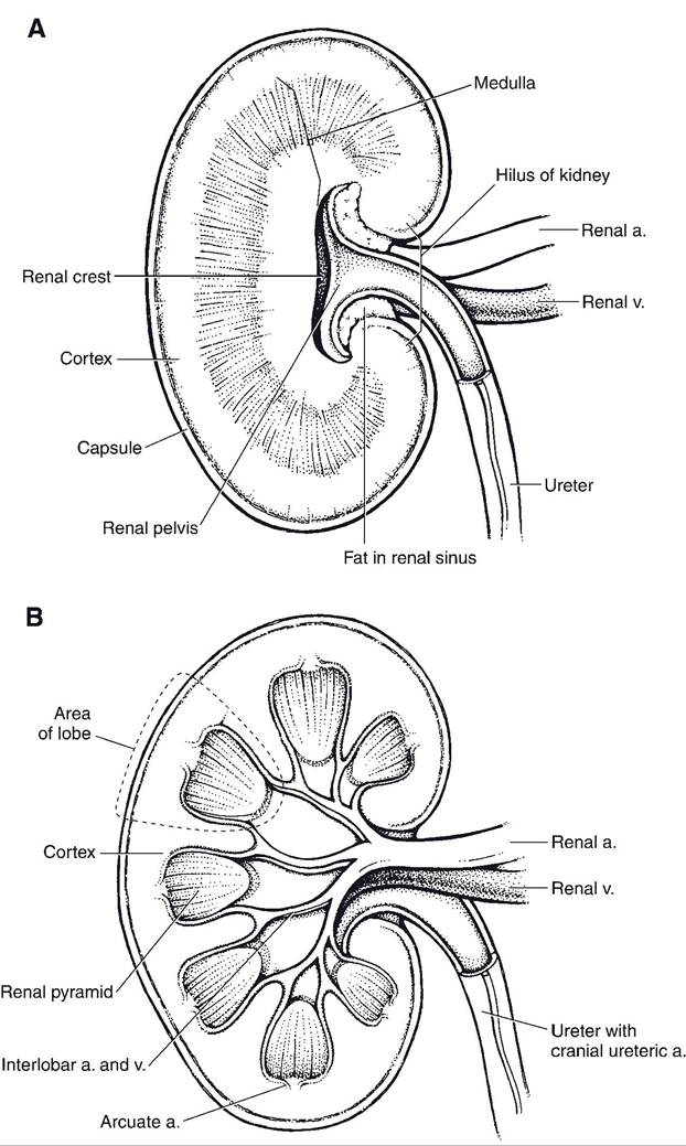
Figure 23-3. Kidney of the canid cut on the median plane (A) and a sagittal section off midline (B). The small ruminant kidney appears similar to this canine kidney. (Reprinted with permission of Wiley-Blackwell from Smith B.J. Canine Anatomy. Philadelphia: Lippincott Williams & Wilkins, 1999.)
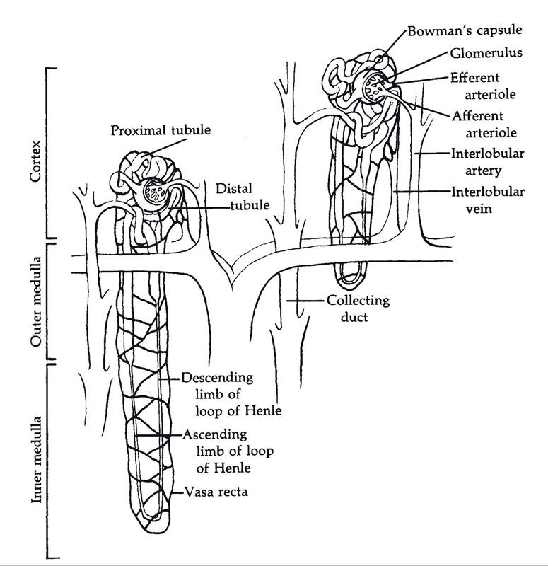
Figure 23-4. Two mammalian nephrons and the microcirculation associated with them. (Reprinted with permission of Wiley-Blackwell from Reece W.O. Physiology of Domestic Animals. 2nd ed. Baltimore: Williams & Wilkins, 1997.)
Arcuate veins drain blood from both the cortex and medulla, pass through the medulla as interlobar veins, and enter the renal veins, which emerge from the renal hilus to empty into the caudal vena cava. Lymph drains from the kidney to the renal lymph nodes.
Sympathetic nerves are the primary innervation of the kidneys. These derive from the celiacomesenteric plexus and innervate blood vessels and renal tubules.