Ureters, Urinary Bladder, and Urethra
The ureter is a muscular tube that conveys urine from the kidney to the urinary bladder. The smooth muscle of the ureter undergoes peristaltic waves of contraction that encourage the flow of urine to the urinary bladder.
Each ureter originates at the renal pelvis (or the major calices of the bovine kidney) and empties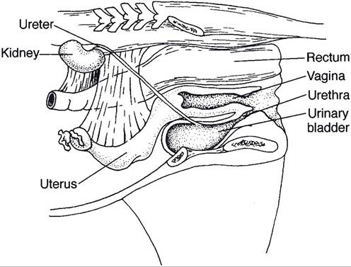
Figure 23-5. Kidneys, ureters, and urinary bladder in situ in the small ruminant. (Reprinted with permission of Wiley-Blackwell from Reece W.O. Physiology of Domestic Animals. 2nd ed. Baltimore: Williams & Wilkins, 1997.)
into the urinary bladder near its neck at the trigone (Fig. 23-5). The manner in which the ureter passes obliquely through the wall of the urinary bladder creates a valve to prevent reflux flow of urine to the kidney.
The urinary bladder is a hollow muscular organ that varies in size and position with the amount of urine it contains. The empty, contracted bladder is a thick-walled, piriform organ on the floor of the pelvic cavity. As it fills with urine, its wall thins, and it enlarges craniad toward and then into the abdominal cavity.
The neck of the bladder is continuous with the urethra caudally. The smooth muscle of the urinary bladder wall is arranged in three sheets; at the neck of the bladder these may form a smooth muscle sphincter that controls passage of urine into the urethra, although the precise role of the intramural muscle of the urinary bladder in creating a true sphincter is debated. The pelvic urethra extends from the urinary bladder across the floor of the pelvic canal to the ischial arch. in female animals, it opens onto the floor of the vaginal vestibule. in the male animal, it receives the ductus deferens and ducts from the accessory sex glands, then passes through the penis as the penile urethra. in both sexes, the pelvic urethra is surrounded by a true sphincter, the striated skeletal m. urethralis, over which the animal exercises voluntary control. The m. urethralis is innervated by the pudendal nerve.
The pelvis, ureter, bladder, and urethra are all lined with transitional epithelium. This epithelial lining is useful in these areas, where considerable distension of the lumen may occur. When these organs are empty, the lumen is small, the walls are thick, and the lining epithelial cells are piled deeply to form many layers. However, when the organs are distended, the lumen is enlarged, the walls are thinner, and a transition to a much lower stratification of the lining occurs (Fig. 23-6). The urethra, like the urinary bladder, is lined with transitional epithelium; this changes to the typical stratified squamous epithelium of mucous membranes at or near the urethral orifice at the tip of the penis of male animals or at its junction with the vestibule of the vagina in the female animal.