ANIMAL EXPERIMENTS
Studying the physiology of organs, organ systems, and the organism as a whole under both natural and experimental situations is the primary goal of the physiology labs. Research is consistently conducted on living subjects or isolated, functional organs.
Sufficient physiological conditions ought to be supplied in both situations. Instrumental measurement and recording enhance and support visual observation. You should document your findings, observations, and conclusions in your notebooks.26.10.1 Anaesthesia and Pain Relief
Several methods exist to prevent pain perception during an operation. Local anaesthesia refers to the reversible inhibition of synaptic transmission or nerve conduction in the operative region. This prevents pain signals from reaching higher centres in the central nervous system. General anaesthesia refers to the total inhibition of synaptic transmission in the nervous system. Adequate anaesthesia means the prevention of pain perception while not interfering with other physiological activities. These characteristics can be addressed by employing agents with a diverse range of actions. Current narcotics achieve anaesthesia levels in the brain, subcortical regions, cerebellum, medulla, and spinal cord, in that sequence (Figure 26.9).
Medication effects can be controlled by carefully choosing the dosage. Cumulative tables provide the anaesthetic dosages per body weight for the most common laboratory animals. The range of efficacy, or the difference between the median lethal dosage (LD50) and the median effective dose (ED50), is also displayed in the tables. (A dose known as the LD50 is one that is fatal for 50% of test participants, whereas the ED50 is a dose that acts in 50% of a population.) The use of the particular medicine is less dangerous the greater this range is. Both the tone of the muscles and the respiratory and circulatory systems are maintained under sufficient anaesthesia (a muscle relaxant is administered to facilitate the release of the latter).
The majority of issues that arise during anaesthesia are breathing-related. It’s also critical to understand that anaesthesia causes a drop in body temperature. Animals undergoing lengthy procedures should therefore be kept warm. The majority of anaesthetics are fat-soluble, meaning that they work by altering the structure of nerve cells’ cell membranes.General anaesthetics are classified as follows:
• volatile liquids (ether, chloroform, halothane)
• gases (nitrous oxide, ethylene, cyclopropane)
• intravenous aesthetic drugs, solutions (barbiturates, pregnanedion, ketamine and urethane)
Volatile or gaseous anaesthetics are commonly utilised in surgery nowadays. In animal research, however, solutions are preferred because they are simple. They can be delivered intraperitoneally (i.p.) or intravenously. The first method is easier, while the second is more effective and allows for more exact drug administration. Depending on the dose and duration of anaesthesia, distinct depths of anaesthesia are distinguished:
• Analgesic time: There is no pain perception and a lowered response threshold.
• Exciting period: Loss of cortical inhibition, absence of consciousness, unconscious escape reactions, speaking sounds, and heightened vegetative functions.
• Period of Tolerance: Surgical procedures are favoured. Subcortical motor centres are blocked, resulting in diminished tone and extinction of somatic reflexes, including the cornea reflex.
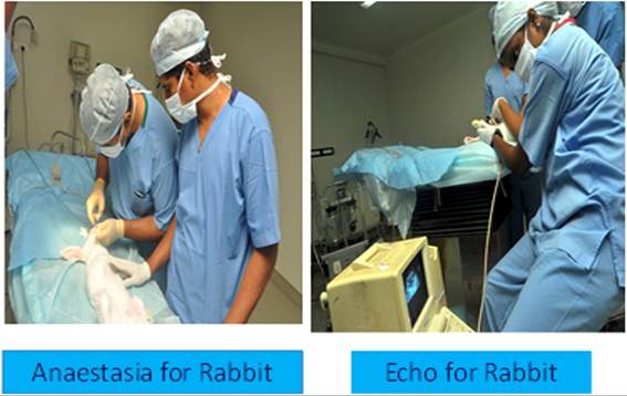
FIGURE 26.9 Myocardial patch for rabbit model at Frontier Mediville.
• A period of respiratory arrest with no medulla activity.
In physiological demonstrations, many anaesthetics are utilised. Short surgeries may be performed under ether anaesthesia (inhalation technique). For longer operations, ketamine with xylazine, nembutal (sodium pentobarbital), or urethane (ethyl carbamate) are employed.
Urethane is only used in operations that end with the animal being sacrificed.
Urethane is a powerful aesthetic for long-term experiments because it influences a wide range of neurotransmissions. Chloralose, commonly in the form of a urethane-chloralose cocktail, is employed in various neurological investigations. It has no effect on reflex or cerebral electrical activity, but its administration is difficult and takes some skill.Procaine or lidocaine local anaesthetics typically block nerve conduction.
26.10.2 Immobilisation of the Animal
In order to execute the procedures precisely and safely, the animals should be attached to the operating board after general anaesthesia or decapitation (frog). Depending on the species and the kind of procedure, there are many ways to fix their bodies. When it comes to frogs, puncturing the limbs to the waxy bottom of the dish can effectively immobilise them. On the other hand, while dealing with rats, the limbs must be secured to the operating board using thread. It is also recommended that the animal’s head be held in place by the upper incisors. When operating with non-anaesthetized (alert) rats (for example, drawing blood samples from the tail vein), the animal is placed in a cone about 5 cm in diameter that only allows access to its tail. Alert rabbits can be investigated in a stock. Rabbits are the least sensitive to pain and the most tolerant to medical procedures of any experimental animal. Before operation under anaesthesia, the rabbits’ forelimbs must be knotted crosswise beneath their backs and their hindlimbs extended apart. The position of the head should also be secured using a thread put between the upper incisors.
26.10.3 Injections
In physiological experiments, parenteral injection of medicines, fluids, and other substances may be required. In these circumstances, agents should be injected. Types of injections include:
26.10.3.1 Subcutaneous (S.C.)
Lift the skin slightly and pierce it in one step. Check for tissue injury and inject the fluid. If there is a big amount of fluid, draw the needle back slightly to prevent it from pooling in this location.
Frogs have lymph sacs under their skin. The injected fluid is rapidly absorbed by them. The lymph sacs are divided by septa.26.10.3.2 Intramuscular (I.M.) Injection
This mode of administration allows for faster and more equal medication absorption. Injections also cause less pain. Injections should be administered immediately and with a forceful movement.
26.10.3.3 Intraperitoneal (I.P.) Injection
For tiny laboratory animals, the abdominal cavity is the recommended place for injections because alternate routes of administration are difficult to implement. Injection involves penetrating the peritoneum. Avoid injuring the internal organs. Drugs delivered intraperitoneally are swiftly absorbed, but their efficacy is significantly influenced by the amount of chyme in the gastrointestinal tract. The doses stated in the table apply to animals that were hungry for 24 hours before to I.P. injection; medication effects cannot be determined in animals that were not starved prior to the experiment.
26.10.3.4 Intravenous (I.V.) Injection
It enables fast and precise administration. Its downside is the difficulty of finding an appropriate vein. Rats and mice use their tail veins. It can be somewhat dilated with warm water. Only a thin needle will yield an effective result. The rabbit’s ear vein is easily localised and can be suitably dilated using a xylene-soaked cotton swab. During this procedure, the rabbit is placed in stocks. Injection of cats and dogs necessitates extensive understanding of the various procedures. It is also possible to execute it in an alert condition with tame animals (especially dogs). Otherwise, it is recommended to use a short-term pre-anaesthesia, such as ether. When administering an intravenous injection, be sure to put the needle parallel to the vein to avoid passing through the vessel wall. Compression of the proximal end of the vein causes a buildup of blood at the injection site, making it much easier to maintain the needle level.
If surgery requires numerous intravenous injections, it is advisable to place a venous cannula in pre-anaesthesia.26.10.4 Preperation of the Animal for Experiment
The animal’s fur disrupts surgical labour since it contaminates the region of operation and, in chronic operations, it can become a source of infection. As a result, it should be shaved off before the surgery. Hairs in rats and mice can be plucked off by hand or with forceps (they regenerate within a few weeks following chronic surgeries). With larger animals (rabbit, cat, or dog), the fur can be shaved with scissors (Cooper’s scissors), but be careful not to hurt the skin. Cut hairs can be removed using a moist cotton swab. A easier alternative is to use a small animal clipper with a cutting head designed to leave just extremely short hairs. Before performing chronic surgeries, the region to be operated on should be painted with iodine or alcohol. This is unnecessary for acute experiments.
26.10.5 Types of Operation
In physiological demonstrations, we usually use acute interventions, which involves killing the animals at the end of the trial. There are other experiments, however, when chronic operations are required. This means that the animals are taken out of anaesthesia and allowed to recover. The most important sorts of operations performed in physiological studies are listed below.
26.10.5.1 In Situ Preperation: Isolated Organ
The organ that will be studied is taken out of its original location and placed within a container. It performs admirably in the synthetic environment of the organ bath. “Straub’s heart preparation” and “The Magnus-type isolated intestinal segment” are two examples of isolated organ preparations.
An organ is removed entirely or is destroyed in part. The effects of an organ’s loss or injury on function are examined. Using this method, the functions of the endocrine glands and specific brain regions can be investigated.
26.10.5.2 Cannulation
It is possible to inject drugs or drain fluids from organ cavities by inserting cannulas.
For instance, an arterial cannula can be used to measure changes in blood pressure, while the cannula placed into the trachea guarantees that the animals under anaesthesia can breathe without problem. Similar to this, gastric and intestinal cannulas can be used to take samples of digestive fluids, while venous cannulas can be used to inject fluids into the body. A ureter cannula can be used to collect urine samples.26.10.5.3 Implantation of Electrodes
Electric stimulators (electrodes) are primarily applied to nerves. The vagus nerve is stimulated to investigate its functions in the heart, lung, and stomach, whereas the ischiadic nerve is stimulated to investigate sensory and motor processes. Electrodes are linked to an appropriate recording apparatus to make recordings.
Naturally, the basic types of operation can be used in combination. A sophisticated physiological experiment may necessitate cannulation, electrode insertion, and organ isolation.
26.10.5.4 Physiological Requirements
To examine animal physiology, appropriate physiological circumstances must be given during the investigations. Three requirements are particularly important. Administered drugs, organ bath, temperature, respiration, heart activity, and blood pressure of animals are all important factors to consider.
“Physiological saline” is the basis for some of the fluids that are injected into the body or used to perfuse isolated organs. Osmotic pressure, ion content, and pH value-wise, they are comparable to the bodily fluids they are meant to replace. Their true makeup may differ according on the experiment’s objectives.
It is necessary to modify the temperature of the injected fluids in addition to their pH value. It is ideal to have a normal body temperature, especially while administering medication intravenously. Controlling the body temperature of animals under anaesthesia is also necessary. A hot water bottle or an electric pad can be used to keep the body temperature of smaller animals at a comfortable level (warming is especially important in the “Complex Physiological” and “Sensory Evoked Potential” tests). Warming up bodily cavities that have been surgically exposed is essential because interior organs are more susceptible to temperature drops. If this isn’t possible, cover the exposed wound with a cotton swab that has been moistened in warm physiological saline.
One possible cause of respiratory failure is anaesthesia. Pre-operative tracheal cannulation can prevent difficulties such as saliva getting into the trachea of the animal lying on its back or the tongue bending back and obstructing the pharynx, which can exacerbate the condition. If this isn’t possible for any reason, give atropine sulphate to lessen the amount of saliva produced.
26.11