ANS: Central components and peripheral components
In the CNS, the main control centres for the ANS are located in the hypothalamus; these can be considered to be autonomic UMNs. The rostral hypothalamus influences the parasympathetic system and the caudal hypothalamus influences the sympathetic system.
Caudally directed fibres synapse in the brain stem and sacral cord (parasympathetic system) or the thoracolumbar cord (sympathetic system). The cerebrum and limbic system can influence but not command the control centres. For example, emotional states, such as aggression or fear, cause piloerection (raising the ‘hackles’). Other parts of the CNS can also influence ANS function as exemplified by olfactory stimulation causing drooling. The hypothalamus integrates autonomic activities associated with temperature regulation, hunger, thirst, sleep, endocrine function and motility of viscera including the gut and urinary bladder. It is also connected to various autonomic brainstem centres that regulate cardiovascular and respiratory function. The cardiovascular centre of the medullary reticular formation can stimulate or depress heart rate. The respiratory centres in the pons and medulla control inspiration and expiration; dysfunction causes abnormal respiration. These areas are regulated by centres in the hypothalamus and the cerebrum.Afferent and efferent fibres of the ANS travel via the spinal and cranial nerves to connect between the CNS and the target organ.
Two-neuron system in the periphery
The ANS comprises two lower motor neurons in series compared with the single LMN in the somatic nervous system. The cell body of the first neuron is in the CNS and it synapses with the second neuron in a peripheral ganglion. The postsynaptic fibre then synapses with the target organ. Presynaptic axons are myelinated and postsynaptic are non-myelinated.
The location of the second neuron is system specific.
For parasympathetic fibres, the ganglion is terminal or intramural; that is it lies close to, or within the wall of, the organ being innervated. Therefore it has a long presynaptic neuron and short postsynaptic neuron.For sympathetic fibres, the ganglion is remote from the organ being innervated. These ganglia are located ventral, or near to the vertebral column in a prevertebral or paravertebral position, respectively. Thus, the sympathetic system has a shorter presynaptic neuron and longer postsynaptic neuron (Fig. 12.2).
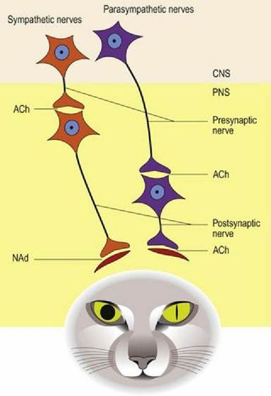
Fig. 12.2 The two neurons that comprise the sympathetic and parasympathetic nervous systems and their neurotransmitters. The effect of each system on the smooth muscles of the mammalian iris is illustrated. ACh = acetylcholine, NAd = noradrenaline.
Note that as the presynaptic nerve can pass through a number of ganglia before synapsing, the terms ‘pre-ganglionic’ and ‘post-ganglionic’ can be misleading. Presynaptic and postsynaptic neurons are the preferred terms.
A summary of the key anatomical features of each system is given in Table 12.1.
Table 12.1 Summary of key anatomical features of each system
| Component | CNS origin | Location of ganglion | Neurotransmitter released |
| Parasympathetic | Cranlosacral | Terminal - dose to organ | Ganglion - acetylcholine Termination - acetylcholine |
| Sympathetic | Thoracolumbar | Distant from organ, e.g. pre/para paravertebral near spinal column | Ganglion - acetylcholine Termination - noradrenaline |