Appendix
The images of the intact and sliced dog brain were obtained from an adult Bull Terrier bitch weighing 33 kg. This brain was perfusion fixed in situ in the head before removal. The transverse slices are photographed from the caudal aspect, except for Figure A19, depicting the caudal midbrain; this was photographed from the rostral aspect.
The photograph of the dorsal aspect of the sheep brainstem (Figure A7) was obtained after curving the brainstem to open the dorsal aspect for visualisation (see inset).
The black and white images are histological sections from a sheep brain. The sections were prepared in 1959, by Dr. A. Palmer, Cambridge, and were derived from interrupted serial sections of sheep brain, embedded in celloidin, cut at 30 μm width and stained for myelin by the Loyez technique, a haematoxylin lake method.
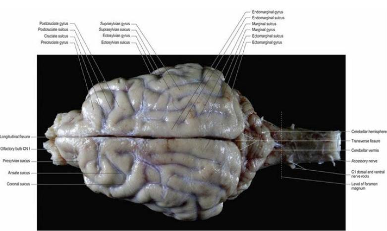
F⅛. A.l Canine brain, dorsal aspect.
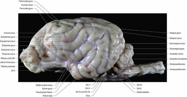
Fig. A.2 Canine brain, lateral aspect.
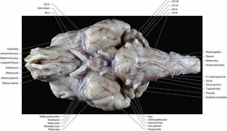
Fig. A.3 Canine brain, ventral aspect.
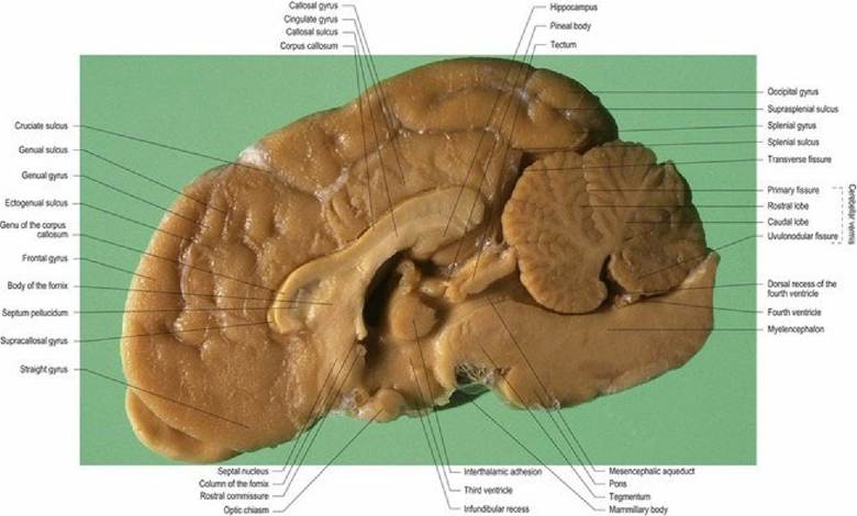
Fig. A.4 Canine brain, median aspect.
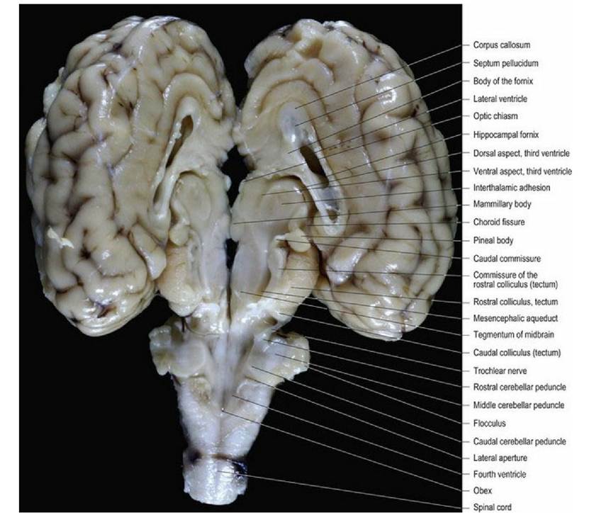
Fig. A.5 Sheep brain, split dorsally to the level of the fourth ventricle. Forebrain and midbrain viewed
from the dorsomedial aspect; medulla oblongata viewed from the dorsal aspect. The bulk of cerebellum has
been removed.
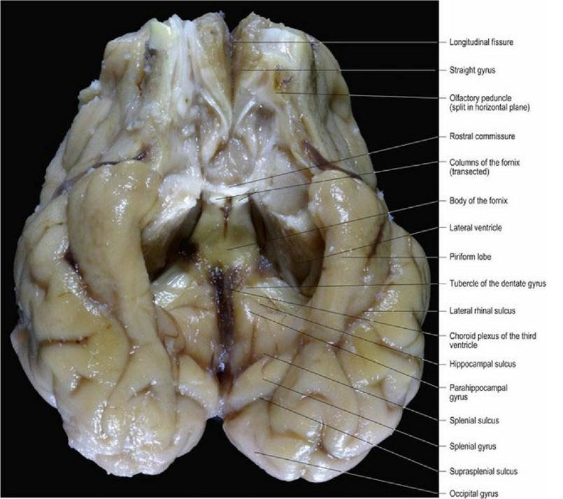
Fig. A.6 Sheep cerebral hemispheres, ventral aspect.
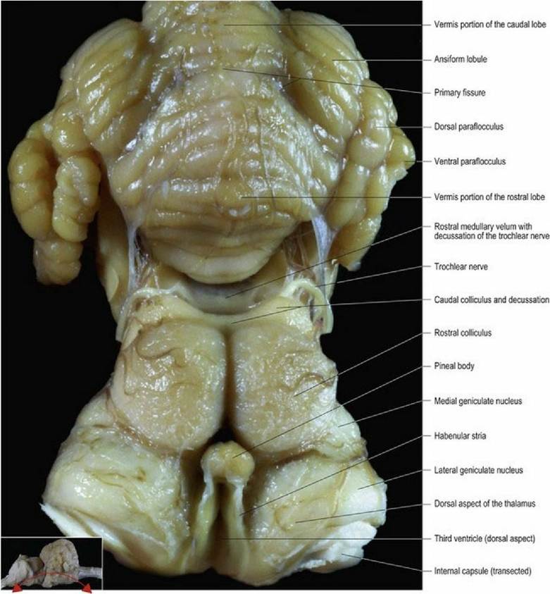
Fig.
A.7 Sheep brainstem, dorsal aspect, cerebral hemispheres have been removed. The brainstem hasbeen curved dorsally (see inset) to better display the anatomical features.
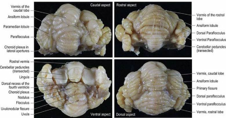
Fig. A.8 Sheep cerebellum, caudal, rostral, ventral and dorsal aspects.
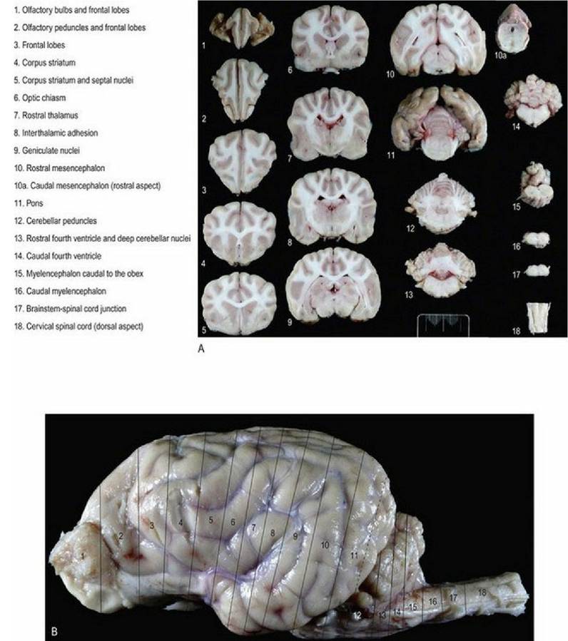
Fig. A.9 (A) Canine brain. The top figure is a general overview of the transverse slices depicted in Figures
A10-25. (B) Canine brain lateral aspect depicting the location of the slices labelled in Fig. A9a. Scale ruler is in millimetres.
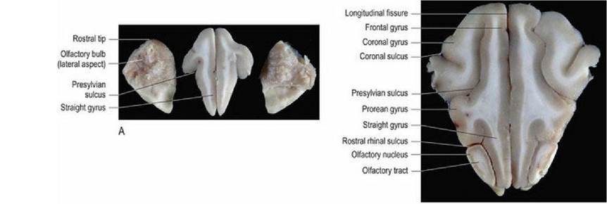
Fig. A.10 (A) Canine brain, transverse slices at the level of the olfactory bulbs. (B) Canine brain
transverse slice through the frontal lobes and olfactory peduncles. (Slices 1 and 2 in Fig. 9a).
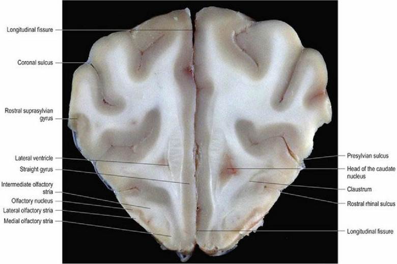
Fig. A.11 Canine brain, transverse slice at the level of the frontal lobes. (Slice 3 in Fig. 9a).
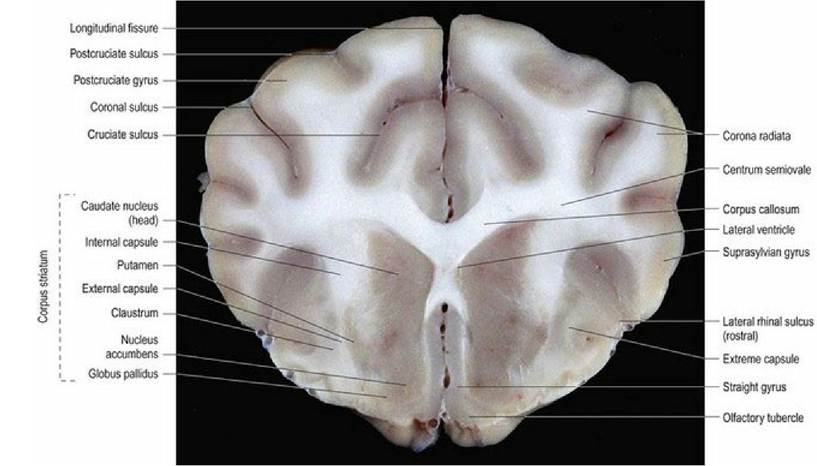
Fig. A.12 Canine brain, transverse slice at the level of the rostral corpus striatum. (Slice 4 in Fig. 9a).
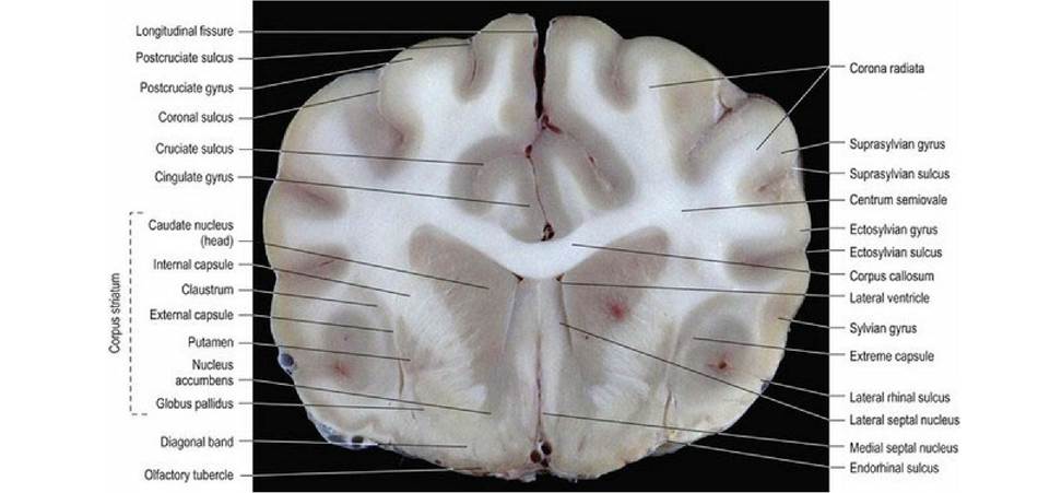
Fig. A.13 Canine brain, transverse slice at the level of the septal nuclei. (Slice 5 in Fig. 9a).
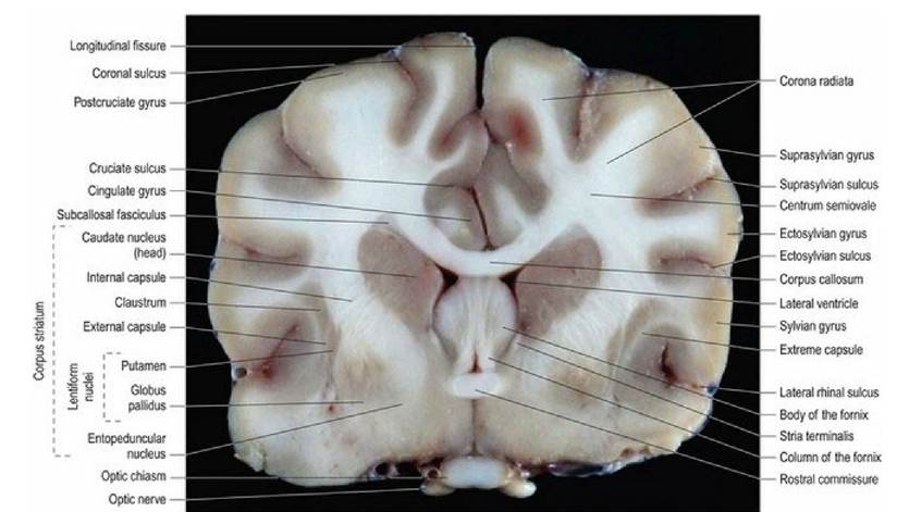
Fig. A.14 Canine brain, transverse slice at the level of the optic chiasm. (Slice 6 in Fig. 9a).
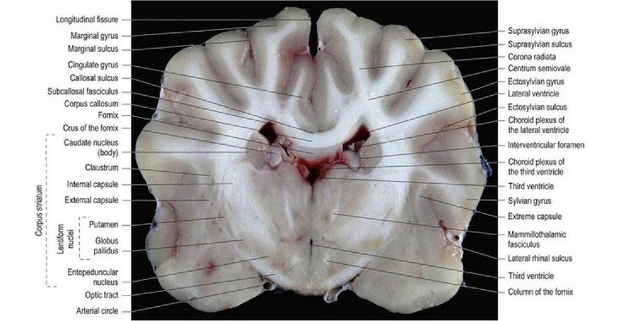
Fig. A.15 Canine brain, transverse slice at the level of the rostral thalamus. (Slice 7 in Fig.
9a).
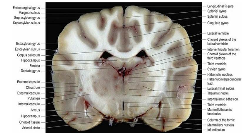
Fig. A.16 Canine brain, transverse slice at the level of the interthalamic adhesion. (Slice 8 in Fig. 9a).
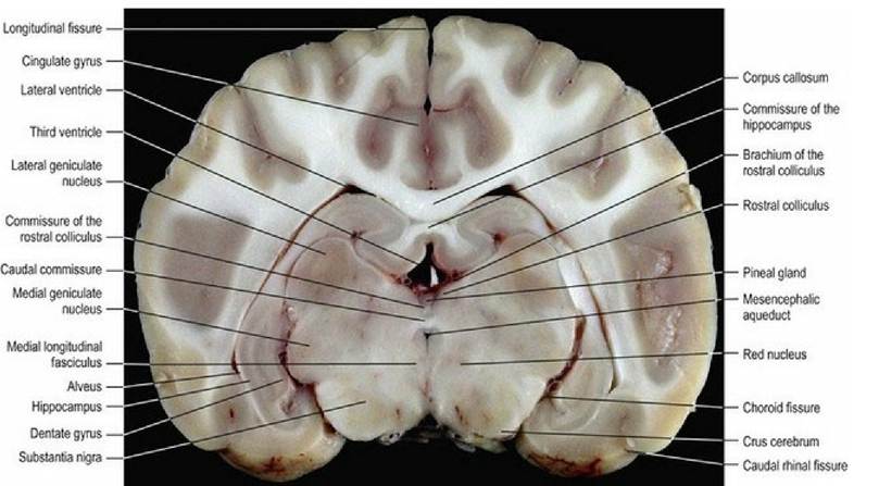
Fig. A.17 Canine brain, transverse slice at the level of the geniculate nuclei. (Slice 9 in Fig. 9a).
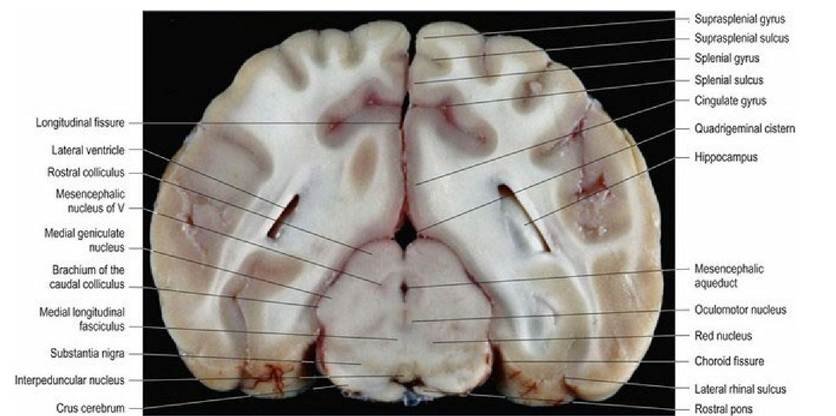
Fig. A.18 Canine brain, transverse slice at the level of the rostral midbrain. (Slice 10 in Fig. 9a).
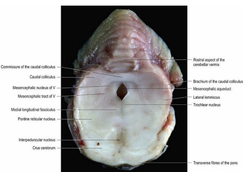
Fig. A.19 Canine brain, transverse slice at the level of the caudal midbrain, viewed from the rostral aspect.
Section is disproportionately enlarged compared with the other transverse sections. (Slice 10a in Fig. 9a).
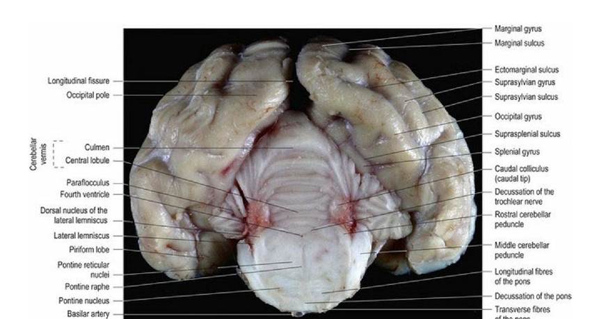
Fig. A.20 Canine brain, transverse slice at the level of pons. (Slice 11 in Fig. 9a).
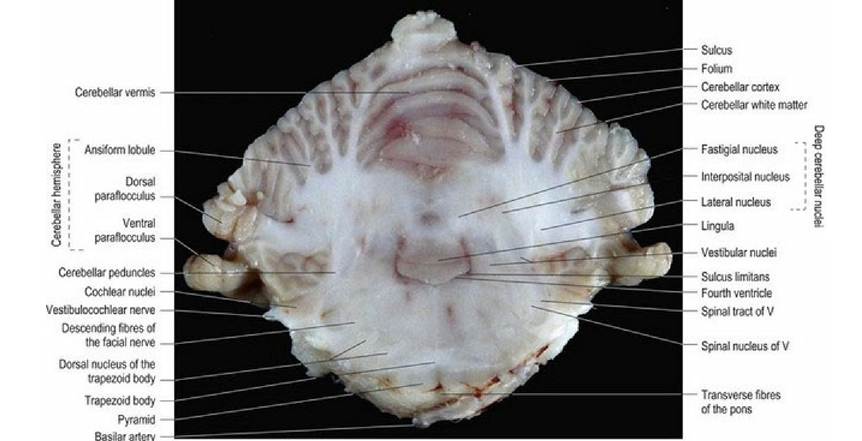
Fig. A.21 Canine brain, transverse slice at the level of the cerebellar peduncles. (Slice 12 in Fig. 9a).
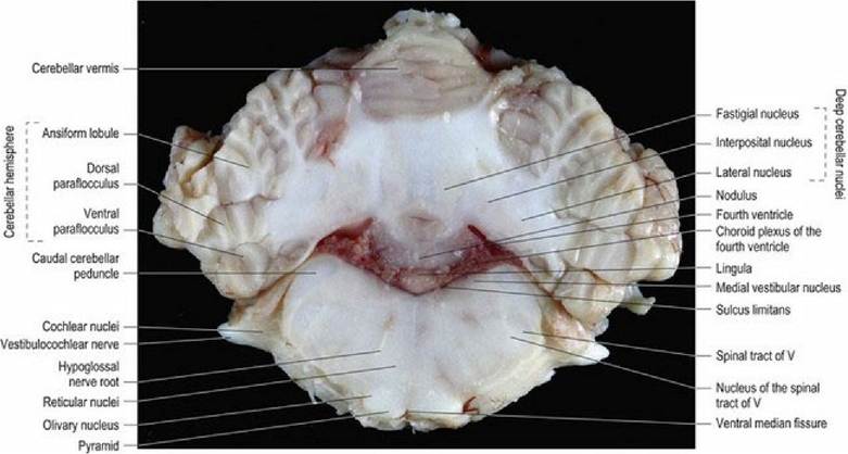
Fig. A.22 Canine brain, transverse slice at the level of the rostral fourth ventricle and deep cerebellar
nuclei. (Slice 13 in Fig. 9a).
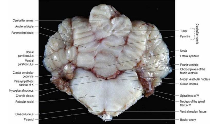
Fig. A.23 Canine brain, transverse slice at the level of the caudal fourth ventricle. (Slice 14 in Fig. 9a).
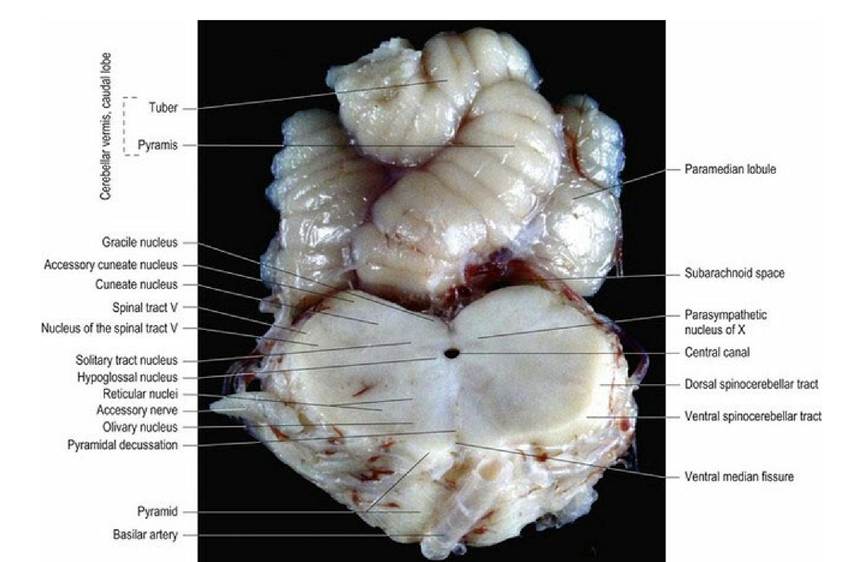
Fig. A.24 Canine brain, transverse slice at the level of the myelencephalon (medulla oblongata) caudal to
the obex.
(Slice 15 in Fig. 9a).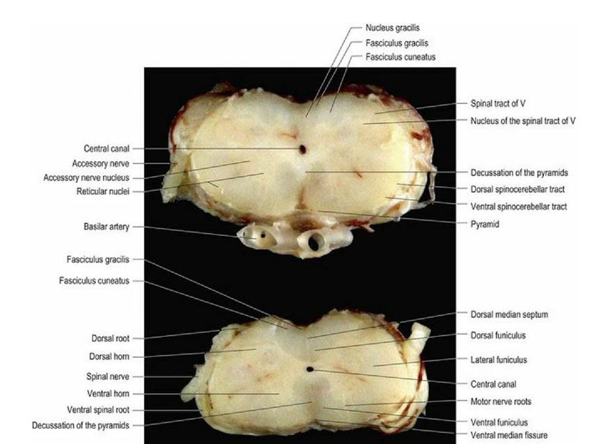
Fig. A.25 Canine brain, transverse slice at the level of the caudal myelencephalon and the brainstem-
spinal cord junction. (Slices 16 and 17 in Fig. 9a).
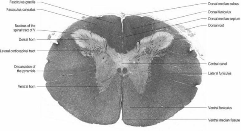
Fig. A.26 Sheep, transverse histological section, C1 spinal cord.
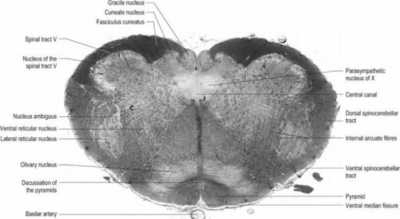
Fig. A.27 Sheep brainstem, transverse histological section, at the level of the brainstem-spinal cord
junction.
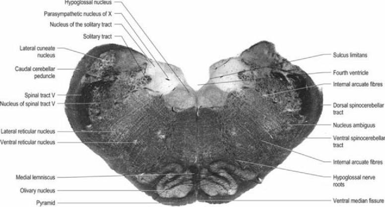
Fig. A.28 Sheep brainstem, transverse histological section, at the level of the caudal medulla oblongata.
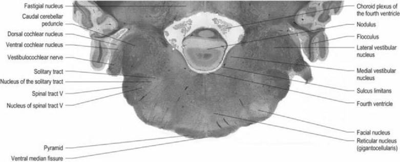
Fig. A.29 Sheep brainstem, transverse histological section, at the level of the rostral medulla oblongata.
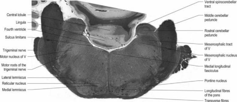
ot the pons Fig. A.30 Sheep brainstem, transverse histological section, at the level of the pons.
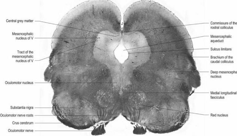
Fig. A.31 Sheep brainstem, transverse histological section, at the level of the rostral mesencephalon.