Arousal
The ascending reticular activating system (ARAS) and related structures are responsible for arousal (consciousness/awareness) (Fig. 11.2). The ARAS is part of the reticular formation and extends from the medulla oblongata to the thalamus.
It determines the level of arousal. It also filters, and prioritises, the plethora of incoming sensory information, for projection to the cerebrum.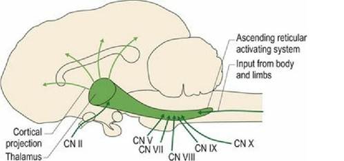
Fig. 11.2 The ascending reticular activating system.
Incoming information in the conscious projection pathways is projected specifically to the defined cortical receiving areas via the thalamus, e.g. tactile information projects to the somatosensory cortex. The ARAS receives collateral axons from these sensory/afferent axons travelling to the diencephalon. This includes exteroceptive, interoceptive and proprioceptive information. The collateral axons synapse in the reticular formation and then the information is projected to the thalamus, from which it is projected diffusely to the entire cerebral cortex. Thus incoming information travelling via the ARAS delivers a generalised ‘wake-up’ call to the cerebral cortices. It keeps the cortex at a general level of alertness ready to receive specific, incoming sensory information. Physiologically, sleep is associated with decreased ARAS activity. The ARAS is thought to be the ‘seat of consciousness’.
Clinically, decreased consciousness can be due to focal lesions involving the reticular formation of the brainstem, or diffuse lesions of the cerebrum (Fig. 11.3). In veterinary medicine, five different levels can be described. Note, the commonly used term ‘depressed’ is a psychological term and is inappropriate:
1. Normal - bright, alert and responsive;
2. Obtunded - dull, tends to fall asleep if left undisturbed, but can be aroused by non-noxious stimuli;
3.
Stuporous - somnolent; requires a noxious stimulus to arouse it;4. Comatose - unconscious; cannot be roused by even a noxious stimulus;
5. Brain dead - no cerebrocortical electrical activity, no brainstem reflex function.
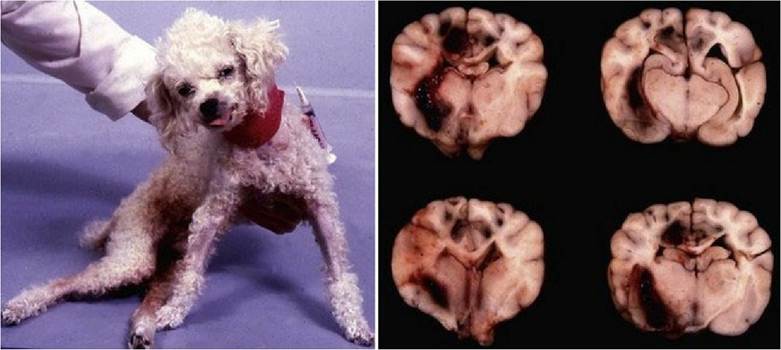
Fig. 11.3 Stuporous dog after head trauma; the dog is being supported. Post-mortem image of transverse brain slices from this dog, depicting the diffuse left-sided haemorrhage and swelling causing compression of the right side of the forebrain.
The brainstem auditory evoked response test (see Fig. 10.18) and brainstem-based cranial nerve reflexes such as the vestibulo-ocular reflex can be used to assess how much brainstem function is still present in comatose animals.
Central to the control of arousal as well as sleep is the neurotransmitter hypocretin (orexin). Hypocretin neurons in the lateral hypothalamus project to areas involved in the sleep-wake cycle, including the ARAS. Hypocretin promotes wakefulness. Hypocretin neurons inhibit the rapid eye movement (REM) stage of sleep by activating brainstem serotonergic and noradrenergic brainstem ‘REM-off neurons, and reduce the activity of pontine cholinergic ‘REM-on’ neurons. The sleep disorder narcolepsy has been associated with a lack of hypocretin production and, in specific lines of dogs, lack of hypocretin receptors (Fig. 11.4).
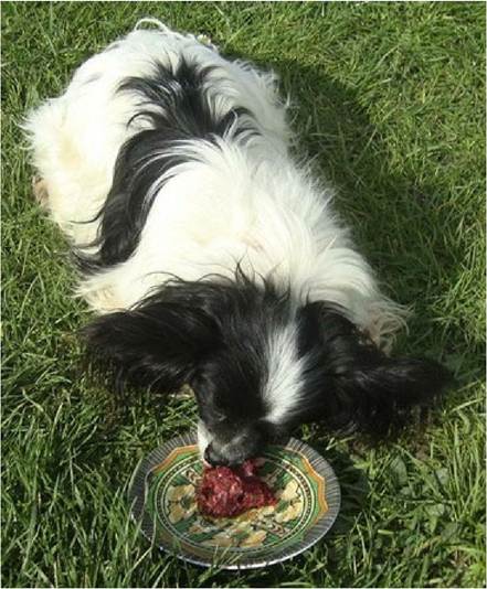
Fig. 11.4 Narcoleptic dog. Typically excitement, such as associated with feeding, can trigger a narcoleptic attack. This dog’s attacks were so frequent that he was having trouble getting sufficient nutrition. Vigorous stroking of the dog would help it to stay awake long enough to eat
(courtesy of Dr. Alison Stickney, IVABS, Massey University).
The ARAS is also thought to act as a filter, or triage system, for incoming information; it evaluates the different inputs in terms of priority. There is a constant barrage of sensory information coming into the brain. The ARAS filters out information that is not considered important at the conscious level and specifically projects important information to the cerebral cortex. Selective filtration helps minimise stimulation by information that is considered to be unimportant at that time, thereby enabling the animal to focus on a particular activity. For example, a cat watching prey may be relatively oblivious to the owner coming up behind it and may be startled when the owner strokes it.