Auditory reflexes, and the brainstem auditory evoked response
Acoustic reflexes
Protective acoustic reflex
When loud, potentially damaging noises occur, input from CN VIII reflexively stimulates CNN V and VII that innervate the tensor tympani and stapedius muscles of the middle ear, respectively.
The tensor tympani muscle attaches to the malleus while the stapedius muscle attaches to the stapes. Contraction of these muscles reduces movement of the ossicles and so limits transduction of sound to the delicate inner ear. The reflex acts bilaterally even after a unilateral stimulus.Olivocochlear reflex
Efferent fibres in CN VIII arise from the olivary nucleus of the medulla oblongata and synapse on the hair cells in the cochlear inhibiting their activation. This reflex may be both protective limiting stimulation of hair cells by sound and also aid in hearing by neutralising the background noise and enhancing the ability to localise the source of a sound.
Reflex head turning
The caudal colliculi receive input from the rostrally directed auditory pathway. The colliculi function in reflexes such as head turning in response to auditory stimuli.
Brainstem auditory evoked response (BAER)
The BAER is an electrical wave form generated by the transmission of nerve impulses along the auditory pathway. It can be used to evaluate the function of components of the auditory pathway from the cochlea to the auditory cortex. In the normal animal, auditory stimuli applied to the external ear result in sequential electrical events occurring in the cochlea, CN VIII, cochlear nuclei and auditory pathways in the brainstem. These evoked potentials can be recorded using surface electrodes placed over the scalp. However, the potential differences of the electrical activity are small and hard to distinguish from the other electrical activity in the brain. Thus headphones are used to deliver hundreds of auditory stimuli (clicks) and the potentials generated are averaged, thereby eliminating background noise.
The resulting wave form has up to seven peaks that are thought to correlate with electrical activity generated in the following structures: I vestibulocochlear nerve, II cochlear nuclei, III nucleus of the trapezoid body, IV lemniscal nuclei, V caudal colliculus, VI medial geniculate nucleus and VII auditory radiations.Lesions that affect peripheral auditory function or the central pathway passing through the brainstem to the forebrain, will alter the wave form. The test was originally used to assess brainstem function, however this has now been replaced by advanced imaging techniques. The BAER are robust however, functioning even if there is no electroencephalogram (EEG) activity, for example after a barbiturate overdose, and assessment of BAER activity can be used to determine whether a patient is ‘brain dead’. More commonly, it is used to assess whether animals, such as Dalmation puppies, are unilaterally or bilaterally deaf due to developmental sensory neural deafness (Fig. 10.18). The latter is a breed-related disease causing degeneration of the stria vascularis, which supplies blood and endolymph to the cochlea; this ultimately leads to degeneration of the hair cells.
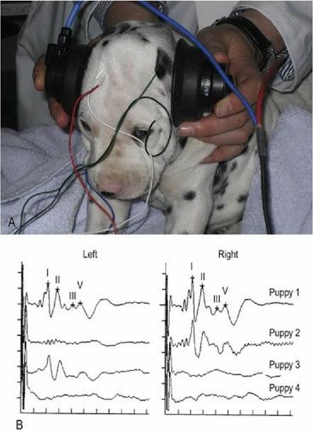
Fig. 10.18 (A) Dalmatian puppy undergoing BAER testing to determine whether it has sensory neural deafness. (B) Brainstem auditory evoked responses from a litter of Dalmatian puppies. Puppy 1 has normal hearing bilaterally, puppies 2 and 3 have impaired hearing in the left and right ears respectively, while puppy
4 has impaired hearing in both ears
(courtesy of Professor J. Mayhew, IVABS, Massey University).
Additionally, hearing may be tested by evaluating Otoacoustic emissions. Auditory stimuli produce a hydromechanical energy wave that travels up the cochlear spiral, stimulating hair cells. This energy wave may be partially re-emitted from the cochlea back into middle ear and result in a sound emitting from the external ear canal. Otoacoustic emissions can be spontaneous or evoked; the latter can be used to test function of the hair cells in the cochlea. This testing has been used for some time in human audiology. It can be used in animals and has been successful in documenting hearing dysfunction in dogs.
Balance and the vestibular system
See Chapter 8.