Audition
Key points
■ The hearing apparatus consists of the outer, middle and inner ears, the cochlear portion of CN VIII, the auditory pathways in the brainstem and the auditory cortex in the forebrain.
■ Sound waves are converted into mechanical vibrations by the tympanic membrane. In the middle ear, the auditory ossicles transmit and amplify the vibrations. The vibrations stimulate pressure waves in the fluid in the inner ear stimulating hair cells in the cochlea. Hair cells transform vibrations into electrical signals that are transmitted by the cochlear nerve to the cochlear nuclei of the brainstem.
■ Output from the cochlear nuclei stimulates auditory reflexes and also travels to the forebrain for the conscious perception of sound.
The hearing apparatus consists of the outer, middle and inner ear, the cochlear portion of the vestibulocochlear nerve (CN VIII) and brainstem auditory pathways in the brainstem. These project to the forebrain and terminate in the auditory cortex in the temporal lobe.
The outer ear in domestic mammals is the receiving device for sound. It is mobile, controlled by muscles at the base and can be turned towards the direction of the sound to improve reception. At the base of the external ear canal is the tympanic membrane or eardrum.
The middle ear is the tympanic cavity. It contains the auditory ossicles and is connected to the nasopharynx via the auditory tube (Eustachian tube in humans). This tube permits pressure to be equalised between the enclosed middle ear cavity and nasopharynx, and hence, the external environment. The three auditory ossicles, located in the middle ear, are the smallest bones in the body. From lateral to medial they are the malleus, incus and stapes. They form synovial articulations with each other. The malleus contacts the tympanic membrane. The stapes contacts the vestibular window and thereby interfaces with the fluid-filled inner ear (Fig.
10.15). The stapedius and tensor tympani muscles connect to the stapes and the malleus and function in protective acoustic reflexes (see next section).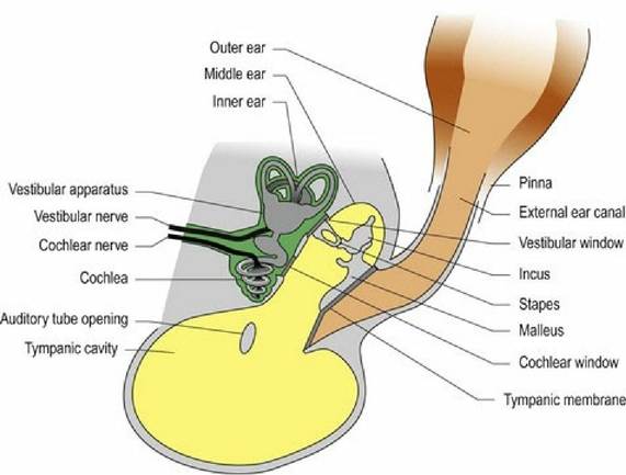
Fig. 10.15 The canine ear.
The inner ear houses the receptors for hearing in the cochlea, and for balance in the vestibular apparatus (see Chapter 8). The inner ear consists of a series of canals and cavities, forming the bony labyrinth. Membrane-bound ducts line the bony labyrinth forming the membranous labyrinth. Between the bone and membrane is fluid called perilymph, while endolymph fills the membranous labyrinth.
The membranous structures of the inner ear are in communication with the middle ear via membrane- covered vestibular (oval) and cochlear (round) windows.
The cochlea is a hollow, snail-shell-like spiral of bone with a central axis, the modiolus (Fig. 10.16). The cavity of the cochlea spirals around the modiolus and membranes divide it into three longitudinal regions creating the scala vestibuli, the central cochlear duct and scala tympani. Perilymph is found in the scala vestibuli and tympani, and endolymph in the cochlear duct. The latter is separated from the scala tympani by the spiral basilar membrane on which hair cells are sited forming the spiral organ. Overlying the hair cells is the tectorial membrane.
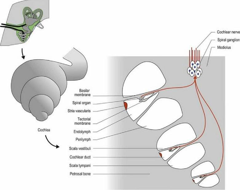
Fig. 10.16 The cochlea. The top image is stylised to depict the spiral nature of the cochlea while the bottom left image is the shape that would be formed by a cast of the bony labyrinth. The bottom right image
is a transverse section through the cochlea.
The terminals of the cochlear portion of the vestibulocochlear nerve synapse with the hair cells. The microvilli of the hair cells contact the overlying tectorial membrane. Sound arriving at the external ear causes the tympanic membrane to vibrate.
This vibration is amplified and carried across the middle ear by the auditory ossicles and results in pressure waves in the fluid of the inner ear. These waves enter the bony cochlea causing vibration of the basilar membrane, which makes the microvilli rub against the tectorial membrane. Deflection of the microvilli on the hair cells results in ion channels opening in the base of the hair cell and depolarisation of the associated nerve endings, generating a nerve impulse.The composition of the basilar membrane varies along its length, so that specific portions only will vibrate in response to a specific frequency of sound. High-frequency sounds stimulate the basilar membrane at the base of the cochlear, while low-frequency sounds stimulate at the apex. Thus, there is frequencydependent stimulation of specific fibres within the cochlear nerve and this is translated into pitch perception in the auditory cortex. Different species can perceive different pitches with dogs hearing frequencies in the range of 67-45 000 Hz, while horses can detect 55-33 500 Hz and humans 64-23 000 Hz. The cochlear window acts as a dampener for excess pressure waves within the cochlea dispersing them into the middle ear cavity.
Audition, the conscious perception of sound, shares the same three-stage system that is the template for most sensory systems. The axons of the first neuron in the pathway form the cochlear portion of the vestibulocochlear nerve (CN VIII) and have their nerve cell bodies located in the spiral ganglion inside the cochlea. The cochlear nerve synapses with the neurons in the dorsal and ventral cochlear nuclei located on the dorsolateral aspect of the rostral medulla oblongata (Figs. A21, A22, A29). Efferent axons from the cochlear nuclei travel rostrally, in the lateral lemniscus, to synapse with the third neuron in the medial geniculate nucleus of the diencephalon (metathalamus). The output from this nucleus travels, via the internal capsule, to the auditory cortex in the temporal lobe for conscious perception of sound (see Fig. 4.13). The pathway is bilaterally represented.
In addition to this simplified three-neuron pathway many fibres synapse in other nuclei as part of the auditory pathway, or for acoustic reflexes. This includes the nuclei of the trapezoid body (caudal border of the pons), the lateral lemniscus (pons and midbrain) and the caudal colliculus of the midbrain (Figs. 10.17, A3, A7, A19).
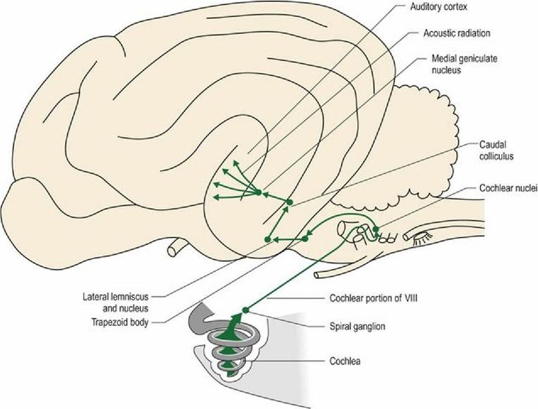
Fig. 10.17 Neural projections from the spiral ganglion to the auditory cortex.
Synapses in the caudal colliculus and connections with the tectonuclear/tectospinal pathways, as described for visual reflexes, are used for reflex eye and head movement in response to sound.