Autonomic Nervous System
The ANS is the part of the nervous system that regulates activity in viscera and other structures not normally under voluntary control (Fig. 916). The common representation of the ANS as a motor subdivision of the peripheral nervous system ignores the facts that (1) sensory fibers from viscera make up a large proportion of the fibers in autonomic nerves and (2) some CNS tracts and nuclei integrate and control visceral activity.
Nonetheless, for purposes of this introduction, we consider only the peripheral motor components of the ANS. These are the nerves that influence activity in smooth muscle, cardiac muscle, and glands.In motor nerves to voluntary muscle, the cell bodies of neurons directly innervating the target are found in the gray matter of the CNS, and the telodendria of these neurons make direct contact with the target. Motor nerves of the ANS, in contrast, consist of a series of two neurons. The first has its cell body in the CNS, and its axon extends into the periphery, where it synapses on the cell body of a second neuron. It is the axon of the second neuron that contacts the visceral target. Because of this two- neuron arrangement, autonomic nerves are characterized by the presence of autonomic ganglia, peripheral collections of the cell bodies of the second neurons. using the autonomic ganglion as a point of reference, the first neuron is called preganglionic, and the second, postganglionic.
The motor output of the ANS is concerned with homeokinesis, the dynamic process of regulating the internal environment to meet the needs of the organism. As a consequence, the motor limb of the ANS is functionally and anatomically divided into two parts. The sympathetic division of the ANS prepares the organism to meet a stress by producing a combination of physiologic changes that increase available fuel molecules, blood flow to muscle, and cardiac output while simultaneously decreasing digestive processes.
The parasympathetic division of the ANS is in many respects the opposite of the sympathetic division. Parasympathetic activity leads to digestion and storage of fuel molecules and acts to bring the organism to a state of rest. The physiologic effects of the two divisions are covered more completely in Chapter 10.Sympathetic Nervous System
Sympathetic nerve fibers arise from thoracic and lumbar segments of the spinal cord, thus the sympathetic division is sometimes called the thoracolumbar division. Preganglionic sympathetic neurons have their cell bodies in a small lateral horn of the spinal cord gray matter, between dorsal and ventral horns. The myelinated axons of these fibers leave via the ventral root, enter the spinal nerve, and then leave it just outside the intervertebral foramen to join a longitudinal chain of autonomic ganglia. one string of these ganglia lies on each side of the vertebral column. Each receives preganglionic fibers from the spinal nerves only in thoracic and lumbar regions, although the chains themselves extend from the cranial cervical region to the most caudal parts of the vertebral column. The ganglia, together with the nerve fibers that link them longitudinally, are called the sympathetic trunk. The ganglia themselves are most correctly called the ganglia of the sympathetic trunk, although they are also called paravertebral or sympathetic chain ganglia. Cell bodies of many of the postganglionic sympathetic neurons are found here. From the sympathetic trunk ganglia, the unmyelinated axons of postganglionic neurons reach their targets either following spinal nerves or via unique autonomic nerves.
In some cases, preganglionic axons pass through the trunk without synapsing and instead synapse on other sympathetic ganglia outside the sympathetic trunk. This second group, collectively known as prevertebral or collateral ganglia, tends to be associated with the large, unpaired arterial branches of the abdominal aorta, after which they are usually named.
No sympathetic preganglionic cell bodies lie cranial to the thoracic spinal cord, so sympathetic innervation to head structures (e.g., the pupil, sweat glands, salivary glands) arrives at
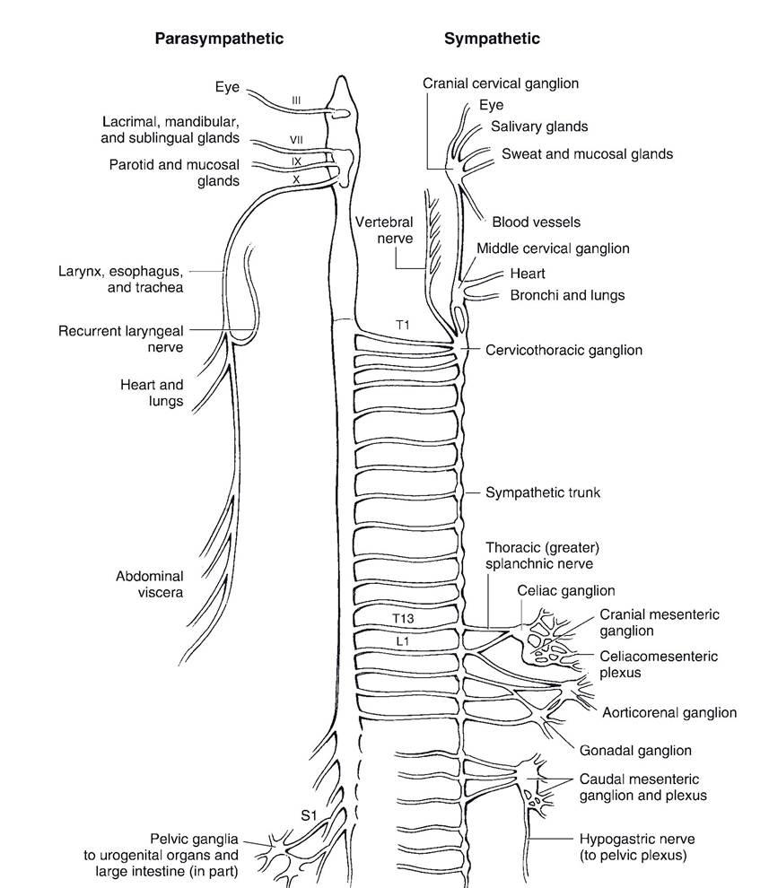
Figure 9-16. The autonomic nervous system. Left) The parasympathetic outflow with cranial nerves III, VII, IX, and X and sacral spinal cord segments carrying parasympathetic fibers. Right) The sympathetic division. Thoracic and cranial lumbar segments make contributions to the sympathetic trunk. These projections are bilateral and are shown on one side only in this drawing for purposes of clarity.
its targets by traveling craniad in right and left bundles of fibers in the ventral neck. These are the cranial continuation of the sympathetic trunks in the thorax. These preganglionic sympathetic fibers are bound in a connective tissue sheath with fibers of each vagus nerve (cranial nerve X); the combined fibers are therefore called the vagosympathetic trunk, readily identified dorsolateral to and parallel with the trachea (windpipe). Sympathetic fibers in the Vagosympathetic trunk synapse in the cranial cervical ganglion, found ventral to the base of the skull, and from this ganglion the postganglionic fibers spread to the glands and smooth muscle of the head.
No sympathetic preganglionic cell bodies exist caudal to the midlumbar region, so sympathetic innervation to pelvic organs (rectum and urogenital organs) arrives via the right and left hypogastric nerves, a continuation of the caudal parts of the sympathetic trunk. The fibers of the hypogastric nerve become admixed with parasympathetic fibers in a diffuse network of autonomic nerves on the lateral surface of the rectum called the pelvic plexus.
The sympathetic innervation to the adrenal gland (see Chapter 11) is unique in that preganglionic sympathetic fibers synapse directly on the chromaffin cells of the adrenal medulla without an intervening ganglion.
Sympathetic stimulation causes this tissue to release catecholamines (epinephrine and norepinephrine) into the bloodstream, producing a widespread, pronounced, and prolonged fight-or-flight response. This is another physiologically important site at which the rapid communication system of the body (the nervous system) is integrated with the slow, more lingering communication system of the body (the endocrine system).Parasympathetic Nervous System
The parasympathetic division of the autonomic nervous system arises from cranial nerves and sacral segments of the spinal cord; for this reason, it is sometimes called the craniosacral division. Fibers of the cranial portion are distributed via four cranial nerves: the oculomotor, facial, glossopharyngeal, and vagus nerves. The first three of these supply parasympathetic fibers to smooth muscle and glands of the head. The vagus nerve supplies parasympathetic fibers to the viscera of the thorax and neck and to nearly all of the abdominal viscera. The distal part of the digestive tract (including the transverse colon and the area caudal to it) and the pelvic viscera are innervated by parasympathetic fibers from the sacral portion of the parasympathetic nervous system. These pelvic fibers intermix with sympathetic nerves to form the pelvic plexus.