Peripheral Nervous System
The PNS includes the nerves and ganglia outside the CNS. Its purpose is to convey sensory information to the brain and spinal cord and to produce movement of muscle and secretion from glands via its motor nerves.
Spinal Nerves
With the exception of cervical and caudal nerves, a pair of spinal nerves (one right and one left) emerges caudal to the vertebra of the same number and name (Fig. 9-13). For example, the first pair of thoracic nerves emerges through the intervertebral foramina between the first and second thoracic vertebrae; the last pair of thoracic nerves emerges through the intervertebral foramina between the last thoracic and first lumbar vertebrae, and the first pair of lumbar nerves emerges through the foramina between the first and second lumbar vertebrae. Thus there are the same number of pairs of thoracic, lumbar, and sacral nerves as there are similar vertebrae.
The first pair of cervical nerves emerges through the lateral vertebral foramina of the atlas and the second pair between the atlas (C1) and axis (C2). Therefore, there are eight pairs of cervical nerves although only seven cervical vertebrae.
Usually, there are fewer pairs of caudal nerves than caudal vertebrae. Five or six pairs are typically seen in domestic ungulates.
Dorsal and ventral roots arise from the spinal cord and fuse, generally close to the intervertebral foramen. At this point, the conjoined sensory fibers of the dorsal root and motor fibers of the ventral root become the spinal nerve, characterized as a mixed nerve, since it has both sensory and motor elements.
Almost as soon as the spinal nerve emerges through the intervertebral foramen, it divides into a dorsal branch and a ventral branch. Both of these branches are mixed nerves, because each contains both sensory and motor fibers.
in general, the dorsal branches of spinal nerves innervate structures (muscles and skin) that are dorsal to the transverse processes of the vertebrae.
The ventral branches supply structures ventral to the transverse processes and most of the thoracic and pelvic limbs.The spinal nerves tend to innervate the region of the body in the area adjacent to where they emerge from the vertebral column. The limbs, however, are supplied with sensory and motor fibers within tangled arrangements of spinal nerves known as plexuses. The regions of the spinal cord supplying the plexuses are visibly greater in diameter because they have more sensory and motor neurons supplying the mass of the limbs. These enlargements are called intumescences.
Brachial Plexus. Each thoracic limb is supplied by a brachial plexus, a network of nerves derived from the last three cervical and first one or two thoracic nerves (Figs. 9-1 and 9-14).
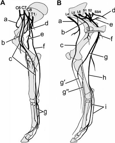
Figure 9-14. A) Nerve supply of thoracic limb of the horse. a, suprascapular n.; b, musculocutaneous n.; c, median n.; d, axillary n.; e, radial n.; f, ulnar n.; g, medial and lateral palmar nn. B) Nerve supply of pelvic limb of the horse. a, femoral n.; b, obturator n.; c, saphenous n.; d, pudendal n.; e, gluteal n.; f, sciatic n.; g, common peroneal n.; g', superficial peroneal n.; g”, deep peroneal n.; h, tibial n.; i, medial and lateral plantar nn.
The spinal cord enlargement associated with the brachial plexus lies primarily in the caudal cervical vertebrae and is consequently described as the cervical intumescence.
The brachial plexus gives rise to specific named nerves that innervate the muscles of the thoracic limb and supply sensation to the same general regions of the skin. Table 9-1 lists the nerves arising from the brachial plexus and the region and muscles supplied by each.
Lumbosacral Plexus. The right and left lumbosacral plexuses supply nerves to the respective pelvic limbs (Figs. 9-1 and 9-14). The lumbosacral plexuses are made up of the ventral branches of the last few lumbar and first two or three sacral nerves.
The visible spinal cord enlargement here is called the lumbar intumescence. The nerves derived from the lumbosacral plexus are described in Table 9-2.Cranial Nerves
Classically, 12 pairs of cranial nerves arising from the brain are described (Fig. 9-15). They
Table 9-1. Nerves of Brachial Plexus
| Nerve | Muscles Innervated | Cutaneous Distribution | |
| Suprascapular | Supraspinatus & infraspinatus | No sensory fibers | |
| Pectorals | Superficial, deep pectoral | No sensory fibers | |
| Subscapular | Subscapularis | No sensory fibers | |
| Musculocutaneous | Biceps brachii Coracobrachialis Brachialis | Medial aspect of antebrachium, carpus; craniomedial aspect of metacarpus | |
| Axillary | Teres minor et major Deltoid | Shoulder region | |
| Radial | Triceps brachii Anconeus Extensor carpi radialis Common and lateral digital extensors Ulnaris lateralis Extensor carpi obliquus Supinator | Craniolateral aspect of antebrachium | |
| Ulnar | Flexor carpi ulnaris Deep digital flexor Intrinsic mm of digit (when present) | Caudal aspect of antibrachium, craniolateral aspect of metacarpus, pastern/foot | |
| Median | Flexor carpi radialis Superficial and deep digital flexor Pronator teres (when present) | Caudal metacarpus, pastern/foot | |
| Thoracodorsal | Latissimus dorsi | No sensory fibers | |
| Lateral thoracic | Cutaneous trunci | No sensory fibers | |
| Table 9-2. Nerves of Lumbosacral Plexus | |||
| Nerve | Muscles Innervated | Cutaneous Distribution | |
| Cranial gluteal | Middle and deep gluteal, tensor fasciae latae | No sensory fibers | |
| Caudal gluteal | Superficial gluteal Parts of middle gluteal, semitendinosus, biceps femoris in horse | No sensory fibers | |
| Femoral | Sartorius Quadriceps femoris Iliopsoas | Medial aspect of thigh | |
| Obturator | Adductor Gracilis Pectineus Obturator externus | No sensory fibers | |
| Sciatic | Semitendinosus et semimembranosus Biceps femoris Obturator internus Gemelli Quadratus femoris | Sensory fibers arise from distal branches (peroneal & tibial nn.) | |
| Peroneal | Tibialis cranialis Extensor digitorum longus et lateralis Peroneus | Dorsal metatarsus and pastern/foot | |
| Tibial | Gastrocnemius Flexor digitorum superficialis et profundus Tibialis caudalis Popliteus | Caudal crus, plantar metatarsus, pastern/foot | |
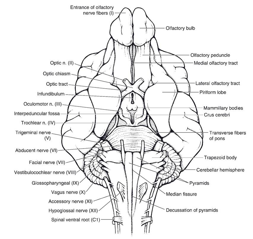
Figure 9-15.
Ventral view of the canine brain. (Reprinted with permission of Wiley-Blackwell from Smith B.J. Canine Anatomy. Philadelphia: Lippincott Williams & Wilkins, 1999.)are designated by Roman numerals, numbered from most rostral (I) to most caudal (XII). With the exception of cranial nerves I (olfactory) and II (optic), the cranial nerves arise from the midbrain, pons, and medulla oblongata and in general resemble ordinary spinal nerves. They do not, however, have discernible dorsal and ventral roots, and some are strictly motor or sensory (as opposed to spinal nerves, which are all mixed nerves).
Cranial nerve II, the optic nerve, only superficially resembles an actual nerve of the PNS. Its fibers are actually a tract of the CNS, invested with meninges and with myelin provided by oligodendrocytes. Features of the 12 cranial nerves are outlined in Table 9-3.
| Table 9-3. Synopsis of Cranial Nerves | ||||
| Number | Name | Type | Arises From | bgcolor=white>Function and Distribution|
| I | Olfactory | Sensory | Olfactory bulb | Olfaction (smell); nasal mucosa |
| II | Optic | Sensory | Diencephalon | Vision; retina |
| III | Oculomotor | Motor | Midbrain | Motor to extraocular eye muscles; parasympathetic innervation to iris sphincter and ciliary muscles |
| IV | Trochlear | Motor | Midbrain | Dorsal oblique muscle of eye |
| V | Trigemina | Mixed | Pons | |
| Ophthalmic division | Sensory | Sensory to eye and dorsal parts of head | ||
| Maxillary division | Sensory | Sensory to maxillary region, nasal cavity, palates, upper teeth | ||
| Mandibular division | Mixed | Sensory to tongue, lower teeth and jaw; Motor to muscles of mastication | ||
| VI | Abducens | Motor | Medulla | Lateral rectus and retractor bulbi muscles of eye |
| VII | Facial | Mixed | Medulla | Sensory (taste) to rostral two-thirds of tongue; parasympathetic to salivary and lacrimal glands; motor to muscles of facial expression |
| VIII | Vestibulocochlear | Sensory | Medulla | Hearing (cochlear division) and sense of acceleration (vestibular division) |
| IX | Glossopharyngeal | Mixed | Medulla | Sensory (taste) to caudal third of tongue; parasympathetic to salivary glands; motor to pharyngeal muscles |
| X | Vagus | Mixed | Medulla | Sensory to pharyngeal, laryngeal mucosa, most viscera; parasympathetic to cervical, thoracic and most abdominal viscera; motor to pharyngeal, laryngeal muscles |
| XI | Accessory | Motor | Medulla & cervical spinal cord | Motor to cervical, shoulder muscles (e.g., trapezius) |
| XII | Hypoglossal | Motor | Medulla | Muscles of tongue |