AVIAN DIGESTION
1. What structures in birds provide for the mechanical breakdown of their ingested food?
2. Do birds have salivary glands and taste buds?
3. Where is the crop located and what is its function?
4.
What are the secretions of the proventriculus?5. What is the function of the gizzard?
6. Do the common domestic birds have gallbladders?
7. What is the most noticeable function of the ceca?
8. What is the most striking feature of colonic motility?
9. What prevents colonic material from entering the ileum during antiperistalsis?
0. What is the function of the bursa of Fabricius?
11. What part of the digestive tract accounts for most of the end products of digestion?
2. How does heat stress interfere with absorption?
3. What is the cloaca?
Differences in the anatomy of the digestive tract were noted among the domestic mammalian species, and although there are general similarities between the domestic avian digestive tracts and those of mammals, there are major differences.
Inasmuch as birds do not have teeth, the mechanical breakdown of their ingested food is accomplished by their beak and by their muscular gizzard. Salivary glands are present in birds and are well developed in those that eat dry foods. Taste buds are located on the tongue and other parts of the mouth as in mammals.
The Digestive Tract
The digestive tract of a turkey is shown in Figure 12-45 and is similar to that of a chicken. The esophagus is divided into precrop and postcrop segments. It is comparatively larger in diameter than in mammals to accommodate the swallowing of large food items that would have been divided in mammals by teeth. Mucous glands are abundant in the esophagus to provide lubrication for food being swallowed. The crop is a dilatation of the esophagus and has a food storage function.
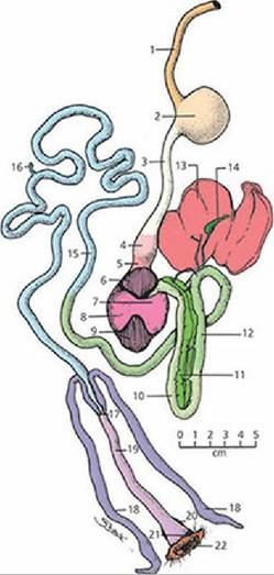
■ FIGURE 12-45 Digestive tract of a turkey.
1, Precrop esophagus; 2, crop; 3, postcrop esophagus; 4, glandular stomach (proventriculus); 5, isthmus; 6-9, muscular stomach (gizzard); 10, proximal duodenum; 11, pancreas; 12, distal duodenum; 13, liver; 14, gallbladder; 15, jejunum; 16, Meckel’s diverticulum (remnant of yolk sac); 17, ileocecocolic junction; 18, ceca; 19, colon; 20, bursa of Fabricius; 21, cloaca; 22, vent. See text for description of the various parts. (From Duke G. Avian digestion. In: Swenson MJ, Reece WO, eds. Dukes’ Physiology of Domestic Animals. 11th edn. Ithaca, NY: Cornell University Press, 1993. Used by permission of the publisher, Cornell University Press)The proventriculus is located between the postcrop esophagus and the gizzard. The gastric secretions HCl and pepsinogen and the mucus are secreted by the proventriculus. Food does not stay in the proventriculus but rather continues into the gizzard, where gastric secretion activity (proteolysis) occurs.
The gizzard is the muscular stomach and is adapted for the mechanical reduction of food that has been ingested.
The small intestine has a well-defined duodenum, with the pancreas located between its loops (as in mammals), but distinction between the jejunum and ileum is not apparent. The yolk sac vestige (Meckel’s diverticulum) is noticeable and is located about midway on the small intestine. One of the liver hepatic ducts proceeds directly to the duodenum; another goes directly to the gallbladder. Gallbladders are present in chickens, turkeys, ducks, and geese. The mucosa of the small intestine is similar to that of mammals, except the villi have well-defined blood capillaries but no central lacteal. The upper ileum is the most important site for absorption of the end products of digested fats, carbohydrates, and proteins. Heat stress and cold stress can be factors affecting absorption. This may be caused by altered mesenteric blood flow (to the intestines), which is decreased about 50% in chickens when the ambient temperature is 37 °C (heat stress).
The large intestine comprises the ceca and the colon. The ceca, which are paired structures, are located at the junction of the small and large intestine. Not all of the food eaten by chickens and turkeys enters the ceca, and the ceca seem to have lesser importance in domestic fowl as compared with wild fowl. The most noticeable function of the ceca is related to the microbial digestion of cellulose. This is of greater importance for the energy needs of some wild species. Urine that has entered the colon from the cloaca may pass into the ceca via antiperistalsis (oral direction). Antiperistalsis is the most striking feature of colonic motility and is believed to occur almost continuously. Because of antiperistalsis, the ceca are filled. A circular muscular ring of the ileum projects into the colon, and its contraction (sphincter-like action) effectively prevents reflux of colonic material into the ileum. In the ceca, the uric acid present in the urine becomes a nitrogen source for the microflora associated with cellulose digestion. Water reabsorption from the refluxed urine is also another important function of the ceca.
The colon is relatively short and links the ileum with the coprodeal compartment of the cloaca; the digestive tract ends with the cloaca, the site that is common to the digestive, reproductive, and urinary systems (Figure 12-46). The coprodeum is the most cranial of the three cloacal compartments followed in order by the urodeum and proctodeum. The three compartments are continuous with each other and are separated only by two annular folds, the coprourodeal and uroproctodeal folds. Urinary and reproductive tracts empty into the urodeum and the proctodeum opens externally through the anus (vent). The bursa of Fabricius is a dorsal diverticulum of the cloaca and is associated with the development of humoral immunity (see Chapter 3). It is an important site for the preprocessing of B lymphocytes.
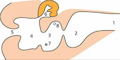
■ FIGURE 12-46 Median section of the cloaca of a 6-month-old female domestic fowl. 1, Colon; 2, coprodeum; 3, urodeum; 4, proctodeum; 5, vent; 6, cloacal bursa; 7, position of oviduct bursa, on left side only; 8, ureteric orifice. (From Reece WO, Trampel DW. Avian digestion. In: Reece WO, ed. Dukes’ Physiology of Domestic Animals, 13th edn. Ames, IA: Wiley-Blackwell, 2015.)
■