AVIAN URINARY SYSTEM
1. Understand the division of the kidneys of birds into lobes and lobules and the structural detail of a lobule.
2. What are the two nephron types associated with avian kidneys?
3.
What is the location of each nephron type within a lobule?4. What is lacking in the reptilian nephron that makes it incapable of concentrating urine?
5. Where are the loops of Henle of the mammalian nephrons located within a lobule?
6. What structures are located in the medullary cone?
7. Would the tubular fluid from reptilian nephrons be exposed to the osmotic gradient in the medullary cone on its exit from the kidney?
8. What is the exit route for ureteral urine?
9. What is the cloaca?
0. Avian kidneys can alternate between reptilian and mammalian nephron types. Which nephron type would promote greater water conservation?
11. Describe the renal portal system.
2. Where does renal portal blood enter the vascular supply that perfuse the peritubular capillary tubules?
3. What is the value of having both arterial blood from mammalian nephrons and portal vein blood from reptilian nephrons perfusing the peritubular capillaries?
4. What is the value of having uric acid precipitated in the peritubular capillary tubules?
L5. What is the principal nitrogenous component of avian urine?
6. What organs in birds are sites for the conversion of ammonia to uric acid?
L7. What is the principal site for the postrenal modification of ureteral urine?
8. What is the extent of urine concentration in birds (osmolality maximum)?
9. What color is bird urine and what is the function of its being mixed with mucus?
:o. How much urine could be produced by a 3-kg hen in a 24-hour period?
There are many similarities between birds and mammals in urine formation and elimination. Also, there are many differences. Similarities include the three phenomena of urine formation, glomerular filtration, tubular reabsorption, and tubular secretion.
Also, birds are able to modify the concentration of ureteral urine so that it may have an osmolality that is above or below that of plasma. Differences between mammals and birds include: in birds, the presence of two major nephron types, the presence of a renal portal system, formation of uric acid instead of urea as the major end product of nitrogen metabolism, and postrenal modification of ureteral urine.Anatomic Features
Avian kidneys are paired retroperitoneal structures that are fitted closely to the bony depressions on the dorsal wall of the fused pelvis. Each kidney has cranial, middle, and caudal lobes (Figure 11-26). Ureters transport urine from the kidneys to the cloaca (mammalian urinary bladder not present). The cloaca is a common collection site, not only for the urinary organs but also for the digestive and reproductive organs. Each lobe has lobules (Figure 11-27) and a lobule gives the appearance of a mushroom, with its cortex corresponding to the cap of the mushroom and the medulla corresponding to the stem.
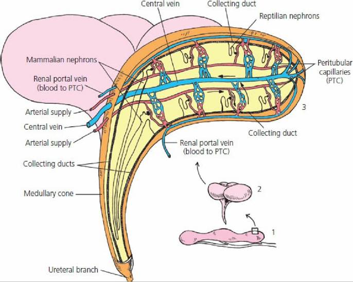
■ FIGURE 11-27 Arrangement of reptilian and mammalian nephrons within a lobule. (1) An avian kidney with its three lobes. (2) A number of lobules from a lobe. (3) The inner structure of a lobule. Reptilian nephrons do not have loops of Henle. Mammalian nephrons are located near the medullary cone and extend their loops of Henle into the cone. The tubular fluid from both nephron types is received by common collecting ducts that also extend into the medullary cone, where it is exposed to ISF concentration gradients similar to mammalian kidneys. All urine from a lobule leaves by a common ureteral branch.
Avian kidneys are characterized by having two nephron types, reptilian and mammalian (Figure 1128). The reptilian-type nephrons lack loops of Henle and are located in the cortex. They are not capable of concentrating urine. Mammalian-type nephrons have well-defined loops of Henle that are grouped into a medullary cone (see Figure 11-27),.the part of the lobule that corresponds to the stem of a mushroom.
Other structures in.the medullary cone are those that would be found in the medulla of a mammalian kidney, the collecting ducts, and vasa recta. The medullary structures enter at the wider cortical end of the cone. The extent of the vasa recta is shown in Figure 11-29. Osmolality of the medullary ISF increases from its beginning near the cortex to the tip of the cone. The osmotic gradient is established by the loops of Henle and is maintained by the vasa recta as in mammalian kidneys, and permits the excretion of urine that has an osmolality greater than that of plasma. All tubular fluid, whether from nephrons of the reptilian or the mammalian type, is exposed to the osmotic gradient because of the exit of.the peritubular collecting ducts through the cone to join the common ureteral branch (see Figure 11-27).
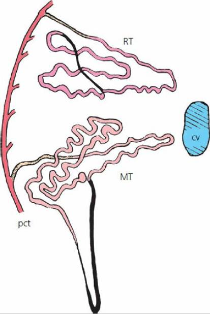
■ FIGURE 11-28 The location of avian reptilian-type (RT) and mammalian-type (MT) nephrons relative to an intralobular central vein (cv) and a perilobular collecting tubule (pct). The intermediate segment of the RT nephron and the nephron loop of the MT nephron are shown in black. The finely stippled areas are beginning collecting tubules. (From Johnson O. Urinary organs. In: King A, McClelland J, eds. Form and Function in Birds. San Diego: Academic Press, 1979.)
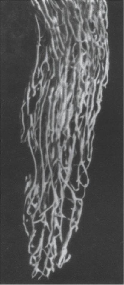
■ FIGURE 11-29 The vasa recta and associated capillary plexus from an avian kidney medullary cone. Microfill injection via ischiadic artery. (From Johnson O. Urinary organs. In: King A, McClelland J, eds. Form and Function in Birds. San Diego: Academic Press, 1979.)
Avian kidneys can alternate between the use of reptilian-type and mammalian-type nephrons, depending on the need for water conservation. Greater use of mammalian-type nephrons would promote greater water conservation. When both nephron types are functional, 25% of the filtrate comes from mammalian-type nephrons and 75% from reptilian-type nephrons.
Renal Portal System
A unique feature of the avian kidney is its renal portal system for part of the blood supply that perfuse the tubules. The renal portal blood is venous blood that comes to the kidney from the hindlimbs via the,external iliac and sciatic veins (Figure 11-30). This venous blood enters the kidney from its periphery, supplying afferent blood to the peritubular capillaries. Within the peritubular capillaries, it is mixed with efferent arteriolar blood coming from the glomeruli (Figure 11-31). The mixture perfuses the peritubular capillary tubules and proceeds to the central vein of the lobule. The renal portal system supplies one-half to two-thirds of the blood to the kidneys. There is a valve, known as a renal portal valve, located at the juncture of the right and left renal veins and their associated iliac veins (see Figure 11-30). Closure of the valve would have the potential of diverting more blood to the renal portal system. Respective adrenergic and cholinergic innervation affect valve closure and opening. The presence of renal portal circulation is of clinical significance when making injections into the hindlimb of birds, because if nephrotoxic substances are introduced the kidney will be subjected to higher concentrations of the drug and permanent damage can result.
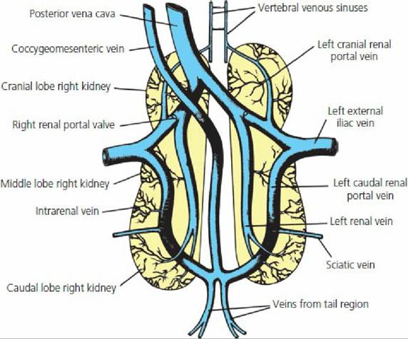
■ FIGURE 11-30 The veins associated with the renal portal system of birds. Blood arrives from the hindlimbs via the external iliac and sciatic veins. Also shown is a renal portal valve. Its closure has potential for diverting more blood to the renal portal system. (From Sturkie PD. Kidneys, extrarenal salt excretion, and urine. In: Sturkie PD, ed. Avian Physiology. 4th edn. New York: Springer-Verlag, 1986.)
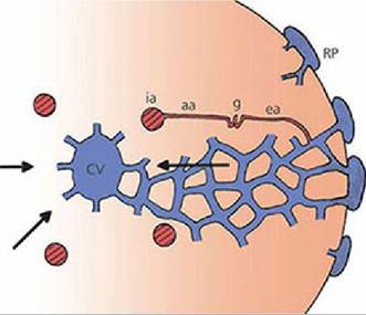
■ FIGURE 11-31 Intralobular blood flow. Intralobular artery (ia) blood supplies afferent arterioles (aa) going to glomeruli (g).
Blood leaving the glomeruli via.efferent arterioles (ea) enters the peritubular capillaries and mixes with blood from branches of the renal portal (RP) veins. Peritubular blood enters the central vein (CV) of each lobule. Arrows indicate the direction of blood flow. (From Johnson O. Urinary organs. In: King A, McClelland J, eds. Form and Function in Birds. San Diego, CA: Academic Press, 1979.)Uric Acid Formation and Excretion
The metabolism of proteins and amino acids results in the production of nitrogenous end products. Among each of the many different kinds of animals, either ammonia, urea, or uric acid accounts for two-thirds or more of the total nitrogen excreted. Accordingly, animals are divided into three groups depending on whether their main nitrogenous excretory product is ammonia, urea, or uric acid. Because ammonia is a very toxic substance, it must be either excreted rapidly or converted to a substance that is less toxic, such as urea or uric acid. Ammonia excretion is encountered only in animals that are entirely aquatic, in which the ammonia can be quickly discharged into their aquatic environment. The urea excreting group is found among mammals and among amphibians.
In reptiles and birds, uric acid is formed instead of urea because these animals develop in eggshells that are impervious to water. The excretion of urea obligates water excretion (because of its effective osmotic pressure) and, because there is only limited water in eggs, it must be conserved. Uric acid reaches a certain concentration and then precipitates. As a precipitate (no effective osmotic pressure), there is no water obligated in its excretion. If urea were excreted it would be necessary to eliminate the liquid urine formed, and this is not possible within eggs.
Just as urea is formed in the liver of mammals from ammonia, so is uric acid formed in the liver of birds from ammonia. The kidneys of birds are also a site for the formation of uric acid. Uric acid precipitates in the peritubular capillary tubules because the increased perfusion of peritubular capillaries that received arterial blood from mammalian nephrons and portal vein blood from reptilian nephrons leads to greater peritubular secretion and consequently greater tubular concentration.
The greater amounts in the tubules exceed uric acid solubility, and it precipitates. Uric acid continues in the perilobular collecting tubules (see Figure 11-28) and collecting ducts (see Figure 11-27) in.its precipitated form and appears in the,urine as a white coagulum. Because uric acid is no longer in solution, it does not contribute to the effective osmotic pressure of the tubular fluid and obligatory water loss is avoided.Concentration of Avian Urine
The permeability of the perilobular collecting tubules and collecting ducts responds to ADH as in mammals. Accordingly, with a need for water conservation, the tubular fluid reaches osmotic equilibrium with the ISF surrounding the tubules, and it becomes hyperosmotic to plasma as collecting tubules and ducts pass through the medullary cone. The hypertonicity of the ISF of the medullary cone is created by NaCl transport from the ascending limbs of the loops of Henle. The maximum concentration of urine that is attainable in birds is much less than for mammals and is about 540 mOsm/kg H2O, which is the concentration of the ISF at the tips of the medullary cones. A maximum urine-to-plasma osmolal ratio would be about 1.58:1.
Modification of Ureteral Urine
After presentation of ureteral urine to the cloaca, there may be retrograde flow into the colon. In the colon, Na+ is reabsorbed and water is reabsorbed by osmosis. The same phenomenon could occur in the ceca if retrograde flow occurred to that level. There is no water reabsorbed from the cloaca even though there may be some Na+ reabsorption.
Urine Characteristics and Flow
Bird urine is cream-colored and contains thick mucus. The precipitated uric acid is mixed with the mucus, whereby the mucus secretion facilitates transport of the precipitate, similar to the mucus in equine urine that facilitates the transport of the carbonates and phosphates that precipitate. Urine flow for hydrated chickens is reported to be about 18 mL/kg/h and for hydrated turkeys it is about 30 mL/kg/h.
■