MAINTENANCE OF ACID-BASE BALANCE
1. As defined, what is an acid and what is a base?
2. A pH of 7.4 is considered normal. What pH would represent a severe acidemia? What pH would represent a severe alkalemia?
3.
What are the three principal chemical buffer systems?4. What are the basic components of the bicarbonate buffer system and the phosphate buffer system?
5. What are the basic groups and the acidic groups of protein buffers?
6. Why is the bicarbonate buffer system considered unique, even though as a buffer system it is considered rather weak?
7. What is the isohydric principal?
8. How is the role of the respiratory system involved in the maintenance of acid-base balance?
9. Study Figures 11-24,11-25, and Figure 11-26 to understand the mechanism for the secretion of H+ ions.
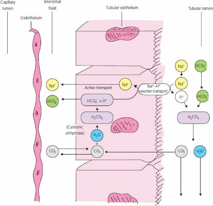
■ FIGURE 11-23 Mechanism for the renal secretion of H+ associated with the bicarbonate buffer system in the tubular fluid.
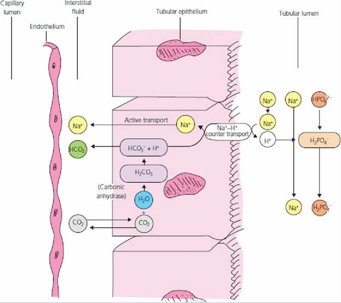
■ FIGURE 11-24 Mechanism for the renal secretion of H+ associated with the phosphate buffer system in the tubular fluid.
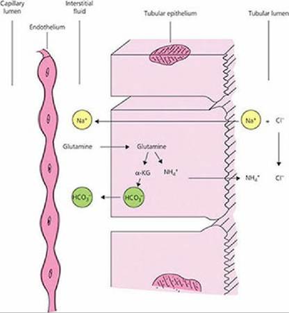
■ FIGURE 11-25 Mechanism for the secretion of H+ associated with the secretion of ammonia by the tubular epithelial cells.
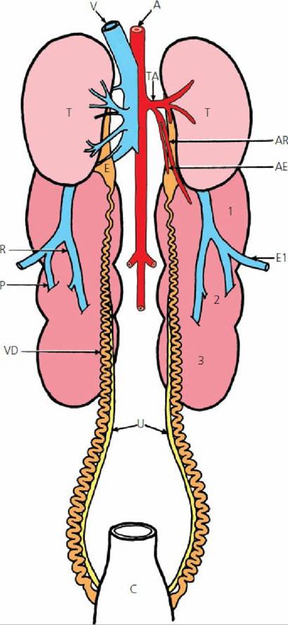
■ FIGURE 11-26 Ventral view of organs and associated structures of the dorsal abdominal cavity of a rooster (male chicken). A, abdominal aorta; AE, epididymal artery; AR, cranial renal artery; C, cloaca; E, epididymis; EI, external iliac vein; P, caudal renal portal vein; R, renal vein; T, testis; TA, testicular artery; U, ureters; V, caudal vena cava; VD, ductus deferens; 1, 2, and 3, cranial, middle, and caudal lobes of the left kidney, respectively.
(From Hodges R. The Histology of the Fowl. New York: Academic Press, 1974.)Under normal conditions, acids or bases are added continuously to the body fluids, either because of their ingestion or as the result of their production in cellular metabolism. In disease, an unusual loss or gain of acid or base may occur as a result of renal insufficiency, insufficient respiratory ventilation, vomiting, or diarrhea. Because of the role of the kidneys and lungs, a study of acid-base balance is important at this point. Three basic mechanisms are involved in the correction of these disturbances: (1) chemical buffering, (2) respiratory adjustment of blood carbon dioxide concentration, and (3) excretion of hydrogen ions or bicarbonate ions by the kidney.
Acids and Bases
The relatively constant [H+] in ECF is the result of a balance between acids and bases. Acids are substances that donate hydrogen ions to a solution; bases are substances that accept and bind hydrogen ions from a solution. A disturbance to this balance occurs when acids or bases are added to or removed from the body fluids. A depression of blood pH to below the normal range is known as
acidemia; a value above the normal pH is called alkalemia. The disturbance caused by the addition of excess acid or the removal of base from ECF is known as acidosis. If it is caused by the addition of excess base or the loss of acid, the disturbance is called alkalosis.
Relationship of pH to H+ Concentration
In their role of regulating the composition of the ECF, the kidneys are important in maintaining a constant hydrogen ion concentration. The pH (negative log of H+ concentration) of the ECF seldom varies from the normal value of about 7.4. A pH change of 0.3 units doubles or halves the H+ concentration. For example, a pH of 7.4 represents a H+ concentration of 40 nEq/L. A pH of 7.1 and 7.7 represents H+ concentrations of 80 and 20 nEq/L, respectively.
In these examples the H+ has doubled or halved from the normal pH of 7.4. A pH of 7.1 represents severe acidemia and a pH of 7.7 represents severe alkalemia.Chemical Buffer Systems
Chemical buffer systems constitute the first line of defense in maintaining constant pH of the ECF. The principal chemical buffer systems are the bicarbonate, phosphate, and protein systems.
The bicarbonate system is represented by NaHCO3 and H2CO3; they react with acid and base as follows:

In Equation 11-1, the basic component of the system reacts with an acid to form a weaker acid and a salt. In Equation 11-2, the weak acid component reacts with a base to form a weaker base and H2O.
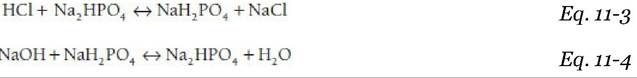
The phosphate buffer system is represented by NaH2PO4 and Na2HPO4. They react similarly to acid and base, respectively:
Proteins act as buffers because their molecules contain a large number of acidic and basic groups. The basic groups (R-NH2) act as buffers by taking up H+ and forming cations (R-NH3+). The acidic groups (R-COOH) act as buffers by losing H+ and forming anions (R-COO-).
Mechanism of H+ Secretion by the Kidneys
The epithelial cells throughout the length of the nephron (except the thin segment of the loop of Henle) secrete H+, but about 85% is secreted by the proximal tubule. The mechanism for the secretion of H+ associated with the bicarbonate buffer system is shown in Figure 11-23. The formation of H2CO3 and the subsequent formation of H+ and HCO3- occurs via the reversible hydration reaction as follows:

The hydration reaction occurs in the cytoplasm of the tubular epithelial cell and is accelerated by the presence of carbonic anhydrase (an enzyme) within the cytoplasm. CO2 in the ECF diffuses freely into the cells.
Increased amounts of CO2 promote more hydration and decreased amounts reduce hydration. After hydration, the H+ formed is secreted into the tubular lumen in exchange for an Na+ (countertransport). The H+ that is secreted combines with the bicarbonate tubular buffer to form H2CO3, which is further dehydrated to the CO2 and H2O that become a part of urine. Dehydration at this location is facilitated by carbonic anhydrase located on the brush border. The HCO3- formed from hydration within the cell diffuses into the ECF, accompanied by the Na+ exchanged for the H+. The ECF loses an H+ and gains an HCO3-. The gain of HCO3- (into the ECF) and the loss of HCO3- (from the tubular fluid) just about balance each other so that pH equilibrium is maintained. When excess hydrogen ions are produced, the phosphates, another tubular buffer, provide for additional exchanges with H+ (Figure 11-24). Figure 11-24 illustrates the independent activity of the phosphate buffer system without regard for the interchanges occurring simultaneously with the bicarbonate buffer system (see Figure 11-23) that continues its buffering activity.The amount of available phosphate remains relatively constant even in acidotic conditions when the kidneys must excrete additional H+ ions. To accommodate the additional H+ ions, a large fraction of excess H+ is excreted in the form of ammonium ions (NH4+). The process begins in the liver with the NH4+ that is an end product of protein metabolism. If the NH4+ is used in the synthesis of urea, H+ ions are released and contribute to the urea load. However, some of the NH4+ is diverted from urea synthesis to the formation of glutamine. The glutamine is carried by the circulation to the tubular epithelium of the kidney and is metabolized to alpha-ketoglutarate (a-KG) and ammonium.
The NH4+ is then secreted into the urine, effectively removing H+ ions from the body. The process of excreting excess H+ ions in the form of ammonium ions is illustrated in Figure 11-25. This process continues in addition to the bicarbonate and phosphate buffering. If acidosis persists, the formation and secretion of ammonia by the tubular epithelial cells increases so that H+ ions can continue to be secreted without lowering the pH of the tubular fluids.Relative Merits of Buffer Systems
The bicarbonate buffer system is rather weak because: (1) the pH of the body fluids is about 7.4 and the pK (negative logarithm of the dissociation constant) of the system is 6.1 (buffering power is greatest when,pH = pK) and (2) the concentration of the buffering elements is not high. The bicarbonate system is unique, however, because it can be adjusted by the respiratory system and the kidneys (i.e., the components are elements of the hydration reaction).
The concentrations of the phosphate buffer components are relatively low in the ECF, but are higher in the intracellular fluids. Accordingly, the phosphate buffer system is more important as an intracellular buffer, not only because of concentration, but also because its pK (6.8) is closer to intracellular pH. Phosphate buffer is also important as a buffer in the kidney tubular fluids when H+ is secreted.
Because of their abundance, the proteins of the body cells, plasma, and hemoglobin (protein of red blood cells) are important chemical buffers. In this regard, hemoglobin is normally the most plentiful chemical buffer in the body. Anemic animals (low hemoglobin concentration) quickly become acidic when they are exerted.
The various buffer systems are not isolated from each other in the body. According to the isohydric principal, any condition causing H+ change results in a balance of all the buffer systems, so they change at the same time (the buffers buffer the buffers).
Role of Respiratory System
Equally important in maintaining acid-base equilibrium in the ECF is the respiratory system (see Chapter 10). During transport from the body cells to the lungs, CO2 diffuses into the erythrocytes and is hydrated under the influence of carbonic anhydrase. The H+ that is formed is buffered and HCO3- diffuses into the plasma. When blood passes through the pulmonary capillaries, the diffusion of CO2 to the alveoli is favored and the hydration reaction is reversed quickly so that H+ is lost from the ECF. Increases in unbuffered H+ cause ventilation of the lungs to increase. Accordingly, the gradient for loss of CO2 into the pulmonary alveoli increases and hydrogen ions are lost at an increased rate. Furthermore, increases in CO2 also increase ventilation so that extra hydrogen ions from increased hydration are lost at the lungs. The role of pulmonary ventilation in regulating H+ concentration can thus clearly be seen.
■