Axial Skeleton
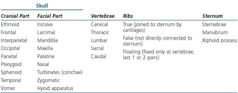
The axial skeleton includes bones on or attached to the midline (axis) of the body and comprise the skull, vertebral column, sternum, and ribs. Table 4-2 lists the bones of the axial skeleton by regions.
Skull
The part of the skeleton within the head is the skull. it protects the brain, supports many of the sense organs, and forms passages for entry
Table 4-2. Bones of the Axial Skeleton
to the digestive and respiratory systems. The skull consists of the cranial part (braincase), which surrounds the brain, and the facial part (Figs. 4-4 and 4-5). The term cranium is sometimes used to denote the entire skull, but more commonly refers only to the braincase, not the facial bones. Most of the observable species differences, as far as the head is concerned, depend on variations in the facial part of the skull.
The caudal and dorsal walls of the cranium are formed by the occipital, parietal, interparietal, and frontal bones. In domestic animals that possess them, the horns have at their core bony projections that arise from the frontal bones. These projections are the cornual processes.
Laterally and ventrally, the walls are formed by the temporal bones, which contain the middle and inner ears, and the sphenoid bone, which supports the brain and pituitary gland. Rostrally, the unpaired ethmoid bone presents numerous openings for passage of the olfactory nerves associated with the sense of smell.
The facial portion of the skull can be divided into orbital, nasal, and oral regions.
The orbit, which means circle, denotes the bony socket that protects the eye. The orbit is surrounded by portions of the frontal, lacrimal, and zygomatic bones. Frontal, zygomatic, and temporal bones all participate in the formation of the prominent zygomatic arch that borders the ventral and caudal parts of the orbit.
The air passages through the nasal part of the skull are bounded dorsally by the nasal bones, laterally by the maxillae and incisive bones, and ventrally by the palatine processes of the maxillae, incisive, and palatine bones. Right and left nasal passages are separated longitudinally by the vomer bone and a cartilaginous and boney septum. Scroll-like conchae (turbinate bones) arise from the lateral walls of the nasal cavity and project into the nasal passages. The conchae are covered with highly vascular mucous membrane that helps warm and humidify the inspired air; conchae in the caudal parts of the nasal cavity feature the olfactory epithelium, which contains the nerve cells specialized to detect odors.
The maxilla and zygomatic bone of the horse feature a sharp ridge, the facial crest, that is readily seen and felt on the lateral aspect of the horse’s head, ventral and rostral to the eye.
Communicating with the nasal cavity are diverticula, known as paranasal sinuses, within some of the bones. The bones that may contain these sinuses include the frontal, maxillary, nasal, sphenoid, and palatine bones. Because it features a diverticulum that extends into the cornual process, the frontal sinus in cattle may be exposed by dehorning mature animals.
The oral (mouth) portion of the skull is roofed by the maxillae and incisive bones and
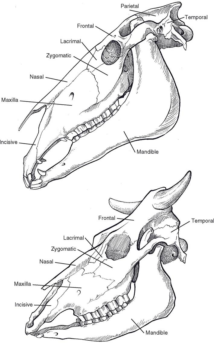
Figure 4-4. Equine (top) and bovine (bottom) skulls, lateral view.

Figure 4-5. Bovine (left) and equine (right) skulls, dorsal view.
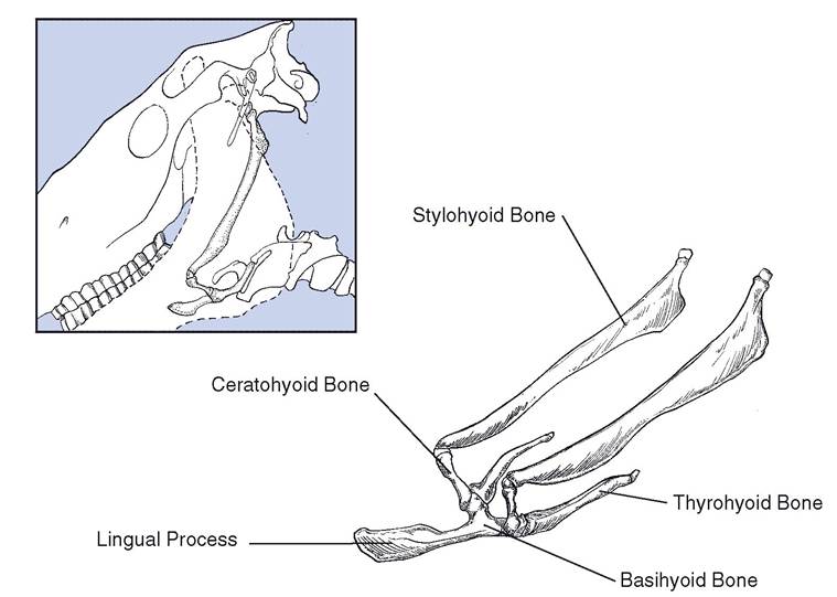
Figure 4-6. Equine hyoid apparatus. The stylohyoid bones articulate with the styloid processes of the skull, and the thyrohyoid bones articulate with the larynx. The prominent lingual process extends into the base of the tongue and affords an attachment site for some lingual muscles.
by the palatine bone.
The maxillae and incisive bones contain the teeth of the upper dental arcade (although the incisive bones lack teeth in ruminants).Ventrolaterally, the mandible completes the oral portion. The mandible pivots on a fossa of the temporal bone just rostral to the opening of the ear. The mandible contains all of the lower teeth and gives attachment to some of the muscles associated with chewing and swallowing.
The hyoid apparatus is a bony framework (Fig. 4-6) that gives support to the pharynx (throat) and provides attachment to some pharyngeal, laryngeal, and lingual muscles. it lies between the right and left portions of the mandible and is attached to the styloid process of each temporal bone.
Vertebral Column
The vertebral column is composed of median unpaired irregular bones called vertebrae. The following letters are typically used to designate the respective regions:
C Cervical vertebrae: neck region T Thoracic: chest
L Lumbar: loin
s sacral: pelvis, fused vertebrae Cd Caudal (coccygeal): tail
A vertebral formula for a given species consists of the letter symbol for each region followed by the number of vertebrae in that region in the given species. Vertebral formulas of common domestic animals and humans are shown in Table 4-3. The parts of a typical vertebra include the body, the arch, and the processes (Fig. 4-7).
The body is a cylindrical mass forming the ventral part of the vertebra and floor of the vertebral foramen.
Dorsally, the arch completes the vertebral foramen, which contains the spinal cord. When vertebrae are placed in series, the adjacent vertebral foramina form the vertebral canal, through which the spinal cord runs longitudinally.
Cranial and caudal articular processes form joints between adjacent vertebrae; in the thoracic region they also form joints with the ribs.
The spinous process projects dorsad from the arch of the vertebra. The spinous processes provide important attachment sites for epaxial muscles.
In the horse, the very tall spinous processes of the first few thoracic vertebrae form a dorsal prominence called the withers.Transverse processes project laterad from the arch.
The intervertebral foramina are formed by the alignment of notches on adjacent vertebrae.
Table 4-3. Vertebral Formulas of Common Domestic Animals and Humans
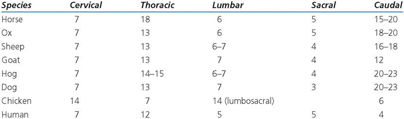
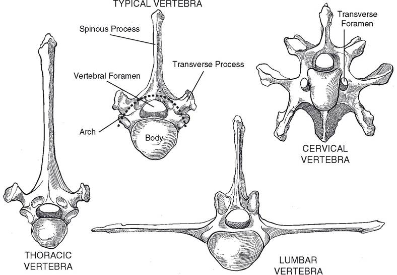
Figure 4-7. Typical vertebrae.
Spinal nerves exit the vertebral canal via the intervertebral foramina on their way to innervating peripheral structures.
The cervical vertebrae have well-developed articular processes to accommodate the large range of motion of the neck. All domestic mammals have seven cervical vertebrae.
The atlas is the first cervical vertebra. The spinous process is absent. The atlas articulates with the occipital condyles of the skull cranially and with the axis caudally.
The axis is the second cervical vertebra. Its spinous process forms a longitudinal sail on its dorsum. The body of the axis features a cranial projection called the dens (for its resemblance to a tooth), which articulates with the atlas in a pivot joint.
The remaining cervical vertebrae are similar to one another, with small spinous processes and rather large transverse and articular processes. With the exception of the last cervical vertebra (C7), each cervical transverse process contains a transverse foramen through which passes the vertebral artery.
Thoracic vertebrae are characterized by well-developed spinous processes and articular facets for the ribs. Costal fovea on the bodies of adjacent thoracic vertebrae form cavities for articulation with the heads of the ribs. Each transverse process also features a fovea for articulation with the tubercle of the rib of the same number as the vertebra.
Lumbar vertebrae have large, flat transverse processes that project laterad.
The spinous processes are similar to those of the last few thoracic vertebrae. The articular processes are more robust than those of the thoracic vertebrae, but not as large as the articular processes in the cervical region. The body and caudal articular processes of the last lumbar vertebra articulate with the sacrum.The sacral vertebrae are fused to form a single wedge-shaped bone, the sacrum, which articulates with the last lumbar vertebra crani- ally, with the first caudal vertebra caudally, and with the wings of the ilia craniolaterally. The intervertebral foramina of the sacrum are represented by dorsal and ventral rows of sacral foramina on dorsal and ventral sides of the sacrum. These foramina, as with other intervertebral foramina, give passage to spinal nerves.
Caudal vertebrae form the bony basis for the tail. Depending on the length of the tail, the number varies considerably from species to species and even within the same species. size of the vertebrae decreases rapidly in a caudal direction, until the last few caudal vertebrae are merely small rods of bone. As animals lack the curved, fused coccyx of human beings, the term coccygeal to describe the vertebrae of the tail is discouraged.
Sternum and Ribs
The sternum forms the ventrum of the bony thorax and gives attachment to the costal cartilages of the ribs as well as providing a bony origin for the pectoral muscles. The cranial extremity of the sternum is the manubrium; the middle portion is the body; and the caudal extremity is the xiphoid process. The sternum consists of individual bones called sternebrae that tend to fuse as age advances. The number of sternebrae (excluding the manubrium and xiphoid from the count) varies with species as follows: pig, four; ruminants and horses, five; and in the dog, six.
The ribs form the lateral walls of the bony thorax. Usually, the number of pairs of ribs is the same as the number of thoracic vertebrae. Rarely, an extra rib or pair of ribs lies either cranial or caudal to the thoracic vertebrae.
A typical rib consists of a shaft, a sternal extremity ventrally, and a vertebral extremity dorsally.Except for the last one or two pairs of ribs, the sternal extremity is connected to the sternum by the costal hyaline cartilage; ribs so attached are called sternal (true) ribs. The vertebral extremity consists of a spherical head connected to the rib by a constricted neck and a tubercle that articulates with the transverse process of a thoracic vertebra. The head articulates with the bodies of two adjacent vertebrae at the costal fovea.
The number of sternal ribs corresponds to the number of sternebrae in the animal. The ribs caudal to the sternal ribs are called asternal (false) ribs because they are not directly connected to the sternum. The costal cartilages at the ventral extremity of most of the asternal ribs overlap and thus indirectly connect the asternal and sternal ribs. Sometimes the last pair or two of ribs have no connection with other ribs at the ventral end. Such ribs are called floating ribs. The spaces between adjacent ribs are the intercostal spaces, numbered to correspond to the number of the rib cranial to the space.