Appendicular Skeleton
The appendicular skeleton is made up of the bones of the limbs. The bones of the thoracic limb are compared to those of the pelvic limb by region in Table 4-4.
Thoracic Limbs
The scapula (shoulder blade) in all animals is a relatively flat triangular bone (Fig.
4-8). The distal portion is its ventral angle, and it forms the only true joint between the scapula and another bone in most domestic animals. Birds and primates possess a clavicle (collarbone), which forms a joint with the scapula, but in most quadrupeds, the clavicle is represented only by the clavicular tendon, a connective tissue band within the brachiocephalicus muscle. In the cat, this tendon may be ossified into a vestigial clavicle. The fused clavicles are called the furcula, or wishbone, in birds. Birds have a coracoid as a separate bone in addition to the scapula and clavicle. The coracoid in humans and domestic mammals has been reduced to the coracoid process (a bony prominence), which protrudes mediad from the scapula near the ventral angle in most species.The lateral face of the scapula has a ridge called the spine extending from the ventral angle to the dorsal border. In carnivores and ruminants, the distal end of the spine is flattened to form the acromion process. The spine divides the lateral face into the supraspinous fossa, which is cranial to the spine, and the infraspinous fossa, which is caudal and ventral to the spine. The costal (medial or deep) face of the scapula gives attachment to some of the muscles that connect the limb to the body.
The humerus (arm bone) is a long bone that varies only in minor details from one animal to another. It has a shaft and two extremities. The proximal end bears a rounded, articular head that participates with the ventral angle of the scapula to form the scapulohumeral (shoulder)
| Table 4-4. Comparison of Bones of Thoracic and Pelvic Limbs | |||
| Thoracic Limb | Pelvic Limb | ||
| Part of Limb | Bones | Part of Limb | Bones |
| Thoracic (shoulder) girdle | Scapula, clavicle, coracoid | Pelvic girdle | Sacrum pelvis: ilium, ischium, pubis |
| Brachium (arm) | Humerus | Thigh | Femur |
| Antebrachium (forearm) | Radius, ulna | Crus (true leg) | Tibia, fibula |
| Carpus (knee) | Carpal bones | Tarsus (hock) | Tarsal bones |
| Metacarpus (cannon and splint bones) | Metacarpal bones | Metatarsus (cannon and splint bones) | Metatarsal bones |
| Phalanges (digit) | Proximal, middle, and distal phalanges Proximal and distal sesamoid bones | Phalanges (digit) | Proximal, middle, and distal phalanges Proximal and distal sesamoid bones |
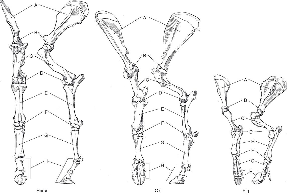
Figure 4-8. Comparative anatomy of the bones of the thoracic limb. A, Scapula; B, scapulohumeral (shoulder joint); C, humerus; D, elbow joint; E, antebrachium (radius & ulna); F, carpus; G, metacarpus; H, digit (phalanges).
joint. The proximal end of the humerus also features a number of irregular tuberosities and tubercles, providing sites of attachment to muscles of the shoulder region. The palpable prominence produced by this end of the humerus is called the point of the shoulder.
The distal end of the humerus forms a spoollike condyle that articulates with the proximal ends of the radius and ulna in the elbow.The radius and ulna are the bones of the antebrachium (forearm). In mammals, the radius is the larger of the two, although in birds it is smaller than the ulna. The radius enters into the elbow joint proximally and the carpus distally. The radius can be felt directly beneath the skin on the medial side of the forearm.
The ulna varies in its degree of development from species to species. The prominent olecranon process (point of the elbow) is found in all mammals proximal and caudal to the elbow joint. This process forms a lever for attachment of the muscles that extend the elbow. In the horse, the proximal portion of the shaft of the ulna is well developed but fused to the radius; the distal ulna is absent. The ox, sheep, goat, and pig each have a complete ulna, but with little or no movement between the ulna and radius. The cat and dog have considerably more movement between these complete bones, but not nearly as much as primates, who can pronate and supinate their hands through the rotation of radius and ulna relative to one another.
The carpus in all animals is a complex region that includes two rows of small bones. This region corresponds to the human wrist, and is frequently, although erroneously, called the “knee” by horsemen. Carpal bones in the proximal row are called (from medial to lateral) radial, intermediate, and ulnar, whereas those in the distal row are numbered 1 to 4 from medial to lateral. In addition, an accessory carpal bone projects caudad from the lateral side of the carpus. The numbering of the carpal bones of the distal row is based on an ancestral four, but among common domestic farm animals only the pig consistently has four carpal bones in this distal row. The first carpal bone of the horse, when present, is small and nonweight bearing. The first carpal is not present in ruminants, and the second and third carpal bones are fused in these species.
The metacarpus is immediately distal to the carpus. In the horse it includes a single large metacarpal (cannon) bone, the base for the third digit (corresponding to the middle finger), and two small metacarpal (splint) bones. The second metacarpal bone is on the medial side, and the fourth is on the lateral side. Trauma to these small bones with consequent excess bone formation results in splints. Splints in horses sometimes produce lameness, but often constitute only a blemish, a disfigurement not usually associated with unsoundness (Figure 4-9).
The cannon bone of the ox and sheep is a fusion of the third and fourth metacarpal bones. A vertical groove on the dorsum of the cannon bone demarcates the embryonic line of fusion.
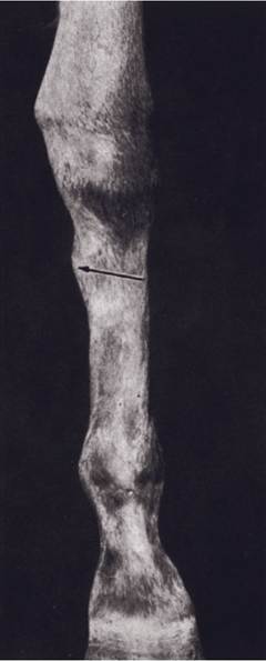
Figure 4-9. Medial splint. (Reprinted with permission of Wiley-Blackwell from Stashak, T.S.: Adams’ Lameness in Horses. 5th ed. Baltimore, Lippincott Williams & Wilkins, 2002.)
The pig has four metacarpal bones. The first is absent; the second and fifth are reduced in size; and the third and fourth bear most of the weight.
The digits number one to five, depending on the species. The horse, having only one digit, literally walks on the tip of the middle finger, or third digit. The digits, like the metacarpal bones, are numbered from one to five from medial to lateral. Each complete digit is made up of three phalanges (proximal phalanx, middle phalanx, and distal phalanx). In the horse, the proximal phalanx is also called the long pastern bone; the middle phalanx corresponds to the short pastern bone; and distal phalanx is also known as the coffin bone. Each digit also includes two proximal sesamoid bones at the palmar aspect of the joint between the third metacarpal bone and proximal phalanx and a distal sesamoid (navicular) bone at the junction of the middle and distal phalanges.
Horsemen refer to the joint between the cannon bone and the proximal phalanx (the metacarpophalangeal joint) as the fetlock.
The portion of the digit between the fetlock and the hoof is the pastern.The ox, sheep, and goat have two principal digits, the third and fourth, whereas the second and fifth digits are represented only by the small dewclaws at the back of the pastern. in the pig the dewclaws are more fully developed as digits (see Fig. 4-8).
Pelvic Limbs
The pelvis consists of a circle of bones by which the pelvic limbs articulate with the vertebral column (Fig. 4-10). Each hemipelvis (half a pelvis) comprises three bones, which are fused to form the os coxae, or pelvic bone (Figure 4-11). These two ossa coxarum are firmly attached to one another at the pelvic symphysis ventrally and are joined to the sacrum of the axial skeleton by two strong sacroiliac joints. The three bones entering into the formation of each ox coxae are the ilium, the ischium, and the pubis. All three of these participate in the formation of the acetabulum of the hip joint.
The ilium is the largest and most dorsal of the pelvic bones. it is irregularly triangular, with the apex at the acetabulum and the base projecting craniodorsad. The medial angle, the tuber sacrale, is close to the sacroiliac joint near the midline. The lateral angle, the tuber coxae, is known as the point of the hip (often called the hook bone by cattlemen). A fracture of the tuber coxae in the horse results in obvious asymmetry in the two points of the hips, as viewed from behind. Horsemen call this condition a knock-down hip.
The broad, flat portion between the tuber coxae and tuber sacrale is the wing of the ilium, and the dorsal margin is the iliac crest. The body of the ilium projects ventrad and caudad between the wing and acetabulum and helps form the lateral wall of the pelvic cavity.
The ischium projects backward and ventrad from the acetabulum, forming much of the floor of the pelvic cavity. The ischium has a large roughened caudal prominence, the tuber ischiadicum (also ischial tuber), commonly called the pin bone in cattle.
The pubis, the smallest of the three pelvic bones, forms the cranial part of the floor of the pelvic cavity. The pubis also enters into the formation of the acetabulum and meets the pubis of the opposite side at the symphysis. The pubis and ischium form the boundaries of the obturator foramen.
The femur (thigh bone) extends from the coxofemoral (hip) joint to the stifle (the joint corresponding to the human knee) (Fig. 4-10). The proximal end of the femur has a nearly spherical head that articulates with the acetabulum of the os coxae to form the hip joint. There are also several roughened prominences, the trochanters, for the attachment of heavy thigh and hip muscles. The shaft of the femur is nearly circular on cross-section and has considerable length. The distal end has two condyles for articulation with the tibia and a trochlea for articulation with the patella, a sesamoid bone embedded in the tendon of insertion of the large quadriceps muscle.
The tibia and fibula are the bones of the true leg (crus), the portion of the pelvic limb between stifle and hock. The tibia, the larger of the two,
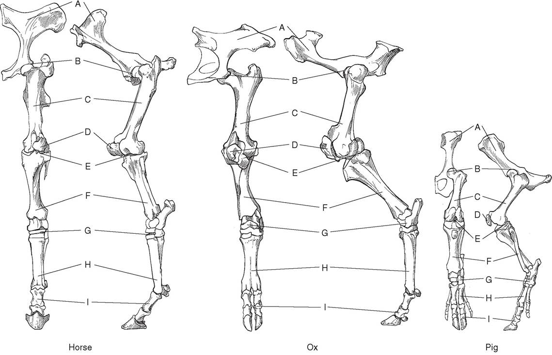
Figure 4-10. Comparative anatomy of the bones of the pelvic limb. A, Pelvis; B, Coxofemoral (hip) joint; C, femur; D, patella; E, stifle (knee) joint; F, crus (tibia & fibula); G, tarsus (hock); H, metatarsus; I, digit (phalanges).
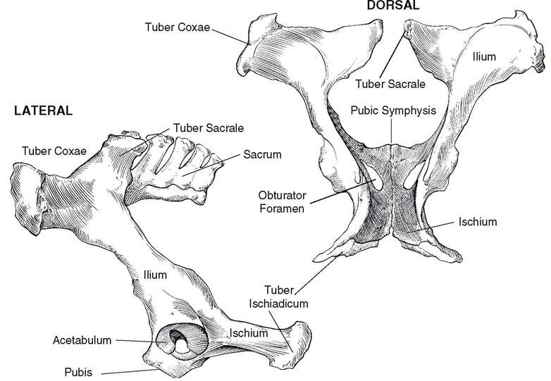
Figure 4-11. The pelvis of the ox. Lateral (left) and dorsal (right) views.
is palpable beneath the skin medially. The fibula, which is much smaller, lies on the lateral side of the leg.
The tibia has an expanded proximal end that participates in the stifle joint. Its shaft is triangular in cross-section. The distal end of the tibia has two concave depressions that form the hinge joint of the hock with the talus (tibiotar- sal bone).
in the dog, pig, and humans, the fibula is a long, thin bone extending from the proximal end of the tibia to the lateral aspect of the hock. The horse has both the proximal end and a portion of the shaft, whereas only a vestige of the proximal end of the fibula is present in domestic ruminants. All domestic species have the distal extremity of the fibula, forming the prominent lateral malleolus of the hock. The lateral malleolus is fused to the tibia in the horse but is a separate small bone articulating with distal tibia and tarsal bones in ruminants.
The tarsus (hock), like the carpus in the thoracic limb is composed of multiple small bones; it corresponds to the human ankle. The proximal row of tarsal bones consists of two large bones. The talus dorsally has two spool-like ridges for articulation with the tibia. The calcaneus projects proximad and caudad to form the point of the hock. The calcaneus, which corresponds to the human heel, acts as a lever for the muscles extending the hock.
in the horse, the central row of tarsal bones is reduced to a single central tarsal bone. The bones of the distal row are numbered 1 to 4 from medial to lateral, with tarsal bones 1 and 2 fused into a single bone. In cattle, tarsal bones 2 and 3 are fused, as are the central and 4th.
The metatarsus and digits of the pelvic limb are similar to the metacarpus and digits of the thoracic limb.