Because Diffusion Is Very Slow, Every Metabolically Active Cell in the Body Must Be Close to a Capillary Carrying Blood by Bulk Flow
To understand more fully how the two types of transport (bulk flow and diffusion) are used in the cardiovascular system, consider the transport of oxygen from the outside air to a neuron in the brain.
With each inspiration, fresh air containing oxygen (O2) moves (by bulk flow) through the trachea, bronchi, and bronchioles and into the alveolar air sacs (Figure 18-2, A). The thin walls separating alveoli contain a meshwork of capillaries (Figure 18-2, B). Blood flowing through these alveolar capillaries passes extremely close (within 1 μm) to the air in the alveoli (Figure 18-2, C). This blood has just returned from the body tissues, where it gave up some of its oxygen. Therefore the concentration of oxygen in alveolar capillary blood is lower than the concentration of oxygen in alveolar air. This concentration difference causes some oxygen to diffuse from the alveolar air into the capillary blood.A large dog has about 300 million alveoli, with a total surface area of about 130 nr (equal to half the surface area of a tennis court). Fhis huge surface area is laced with pulmonary capillaries. Thus, even though only a tiny amount of oxygen diffuses into each pulmonary capillary, the aggregate
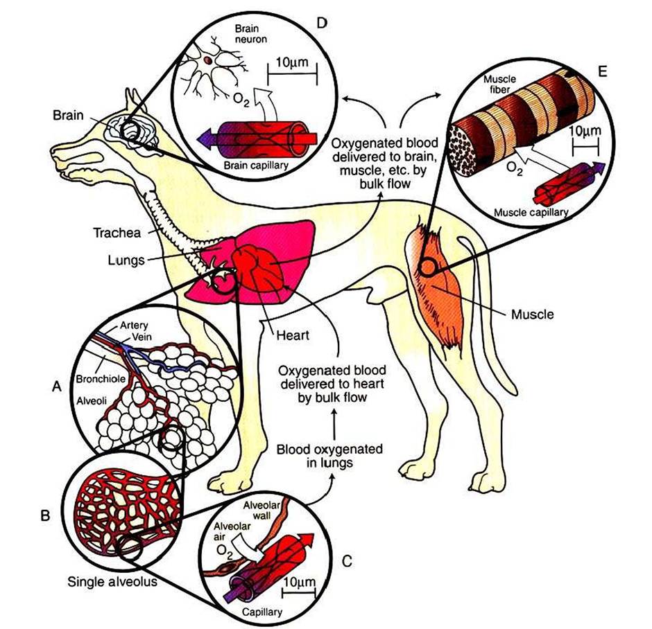
FIGURE 18-2 Oxygen (O2) is transported from the atmosphere to cells throughout the body by a combination of bulk flow and diffusion. First, O2 moves by bulk flow through the airways, from the atmosphere to the alveolar air sacs of the lungs (inset A). From the alveolar air, O2 next diffuses into the blood that is flowing through pulmonary capillaries (insets B and C). Bulk flow of blood carries this O2 to the heart; from there it is delivered by bulk flow into the capillaries of all the body organs (except the lungs).
In the brain (inset D), skeletal muscle (inset E), and other tissues, O2 moves by diffusion from the capillary blood into the interstitial fluid and then into the tissue cells, where it is utilized to support oxidative metabolism. Bulk flow is rapid; it can transport O2 to all parts of the body within a few seconds. Diffusion is slow; it can transport O2 efficiently only over distances less than 100 μm (note distance scales in insets C, D, and E). Oxygenated blood has a bright-red color; deoxygenated blood is darker and bluish red.uptake of oxygen into the pulmonary bloodstream is substantial (typically, 125 mL O2Zminute in a large, resting dog, increasing by a factor of IO or more during strenuous exercise). In summary, both the large surface area and the proximity of alveolar air to capillary blood promote efficient diffusion of oxygen; it takes less than 1 second for the blood in a pulmonary capillary to become oxygenated.
As it leaves the lungs, each IOO mL of oxygenated blood normally carries 20 mL of oxygen. About 1.5% of this oxygen is carried in solution; the other 98.5% is bound to the protein hemoglobin within the erythrocytes (red blood cells). The oxygenated blood moves by bulk flow from the lungs to the heart. The heart pumps this oxygenated blood out into a system of branching arteries, and it is thereby delivered to all parts of the body, including the brain and skeletal muscles (Figure 18-2). Capillaries in the brain bring the oxygenated blood close to each brain neuron (Figure 18-2, D). Metabolic processes within the neurons consume oxygen, so the oxygen concentration inside neurons is low. The gradient of oxygen concentration between the capillary blood (high) and the neurons (low) provides the driving force for oxygen to diffuse first from the blood into the interstitial fluid and then into the neurons.
Each brain neuron must be within about 100 μm of a capillary carrying blood by bulk flow if diffusion is to deliver oxygen rapidly enough to sustain normal metabolism in the neuron.
Diffusional exchange over distances up to 100 μm typically takes only 1 to 5 seconds. If the distance involved were a few millimeters, diffusion would take minutes to occur. Diffusion of oxygen a few centimeters through body fluid would take hours. Therefore, normal life processes require that every Fnetabolically active cell of the body be within about 100 μm of a capillary carrying blood by bulk flow. If this bulk flow is interrupted for any reason, perhaps because of a thrombus (blood clot) in the artery that delivers blood to a particular region of a tissue, that region of tissue becomes ischemic. Again, as stated earlier, ischemia leads to dysfunction, and persistent, severe ischemia leads to infarction and eventually to necrosis. Cerebral infarction causes the condition commonly known as stroke.Figure 18-2, £, shows a capillary carrying bulk flow of blood past a skeletal muscle cell (muscle fiber). Oxygen moves by diffusion from the capillary blood into the muscle interstitial fluid and then into the muscle cell, where it is consumed in the metabolic reactions that provide energy for muscle contraction. The oxygen consumption of a skeletal muscle depends on the severity of its exercise; at a maximum, oxygen consumption may reach levels 40 times greater than the resting level. Because of its tremendous metabolic capacity, muscle tissue has an especially high density of capillaries. In fact, several capillaries are typically arrayed around each skeletal muscle fiber. This arrangement provides more surface area for diffusional exchange than would be possible with a single capillary and also brings the bulk flow of blood extremely close to all parts of each skeletal muscle cell.
Heart muscle, as with skeletal muscle, consumes a large amount of oxygen. Oxygenated blood is carried from the aorta to the heart muscle by a network of branching coronary arteries. This blood moves by bulk flow through capillaries that run close to each cardiac muscle cell.
If a thrombus interrupts the bulk flow of blood in a coronary artery, the heart muscle cells supplied by that artery become ischemic. Ischemia develops even if the cardiac muscle deprived of blood flow lies within a few millimeters of the left ventricular chamber, which is filled with oxygen-rich blood. Oxygen simply cannot diffuse rapidly enough from the ventricular chamber to the ischemic cells to sustain their metabolism. Ischemic cardiac muscle loses its ability to contract forcefully, and cardiac arrhythmias may develop. Severe myocardial ischemia causes a myocardial infarction, or heart attack.Coronary artery disease and cerebrovascular disease are encountered more often in human medicine than in veterinary medicine. In contrast, cardiac disease (dysfunction of the heart muscle or valves, as distinguished from disease of the coronary arteries) is encountered more often in veterinary medicine than in human medicine. Therefore, Chapters 19 to 26 place more emphasis on cardiac physiology than on vascular physiology.