BLOOD SUPPLY
The ventral abdominal wall is supplied by four paired arteries, entering from the sternal and pelvic region, respectively. The cranial superficial epigastric artery branches off the internal thoracic artery, and runs between the abdominal muscles and the skin.
It supplies the region cranial to the level of the umbilicus (it is enlarged in the lactating bitch). The cranial epigastric artery runs deep to the rectus, between it and its sheath. The caudal superficial epigastric artery, a branch of the external pudendal, is distributed subcutaneously and also supplies the prepuce; the caudal epigastric artery arises from the pudendoepigastric trunk and passes forward, first along the lateral border and then on the deep surface of the rectus muscle (see Figures 14-3 and 2-26). Cranial and caudal sets of vessels anastomose (Figure 14-2).The abdominal wall is most safely punctured (paracentesis) a short distance caudolateral to the umbilicus; this site avoids both the fat-filled falciform ligament and risk of injury to a full bladder. The falciform ligament, carrying the round ligament of the liver in its free border, is the remnant of the ventral mesogastrium that conveyed the umbilical vein from the umbilicus to the liver in the fetus. The part adjacent to the liver
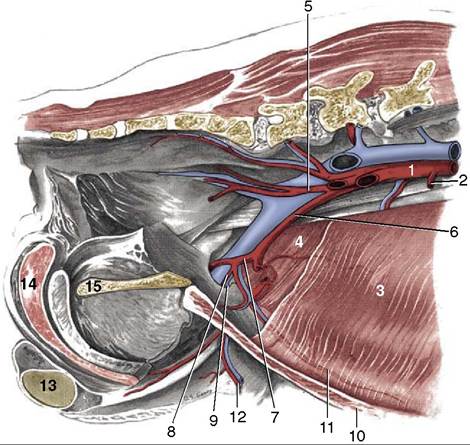
Figure 14-3 Abdominal wall and pelvic canal of the male dog, showing the breakup of the aorta; medial view. 1, Aorta; 2, caudal mesenteric a.; 3, transversus abdominis; 4, internal abdominal oblique m.; 5, internal iliac a.; 6, external iliac a.; 7, deep femoral a.; 8, pudendoepigastric trunk; 9, deep inguinal ring; 10, rectus abdominis m.; 11, caudal epigastric a.; 12, external pudendal a.; 13, left testis; 14, bulb of the penis; 15, pelvic symphysis.
survives, if at all, as a simple peritoneal fold. The blood supply of the falciform ligament arises from along the length of the linea alba. The ligament commonly serves as a major fat storage depot and may become so thickened and enlarged that it complicates the opening and closure of a midline abdominal incision (Figure 14-11), especially in dogs. Part or all of this obstruction may be excised; care must be taken to place a ligature at the cranial end, before the ligament is totally removed.