CARDIOVASCULAR SYSTEM
The heart occupies a relatively large space in the thoracic cavity. It lies in the midline at the level of 2nd-4th intercostal space. The pericardium has two layers: an outer fibrous layer and a thinner serous layer.
Venepuncture
The blood volume of a guinea pig is 70-75 ml/kg of blood. The lateral saphenous and cephalic veins are most accessible but are small. The jugular vein, which lies in the superficial fascia of the ventral neck, can be used for larger blood samples. Compared to the rabbit, rat, and hamster the guinea pig has a longer prothrombin time, so blood clotting on sampling is less of a problem (Sisk 1976).
Unique blood cell features
Lymphocytes
The lymphocyte, which is the predominant peripheral blood cell, is resistant to steroids, as it is in the ferret, man, and monkey. This means that in contrast to the hamster, rabbit, and rat, treatment with steroids does not cause marked
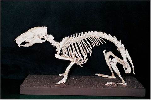
Figure 9.6 • Skeleton of guinea pig (Cavia porcellus).
changes in thymic physiology or peripheral lymphocyte counts (Sisk 1976).
Kurloff cell
This is a unique leukocyte of guinea pigs produced by the thymus under estrogen stimulation. It resembles a lymphocyte but contains oval or round inclusions called Kurloff bodies. These cells are rare in young animals, low in males and related to the estrous cycle in females. They are highest in the female during pregnancy and may play a role in the maternal-fetal barrier. They are also found in high number in the trophoblast region of the placenta (Percy & Barthold 2001; Sisk 1976).
Kurloff cells also appear to have cytotoxic effect on leukemic cells and may explain why spontaneous tumors are not common in guinea pigs (Percy & Barthold 2001).