SKELETAL SYSTEM
Skull
Guinea pigs have large tympanic bullae and prominent zygomatic arches (Fig. 9.7). The mandible, which is the largest bone in the skull, is united rostrally at the mandibular symphysis.
It is roughened laterally for the insertion of the masseter muscle.Axial skeleton
There are 7 cervical, 13-14 thoracic, 6 lumbar, 3-4 sacral and 4-7 caudal vertebrae. There are 13-14 pairs of ribs, with the last 2 being cartilaginous. The first six pairs articulate
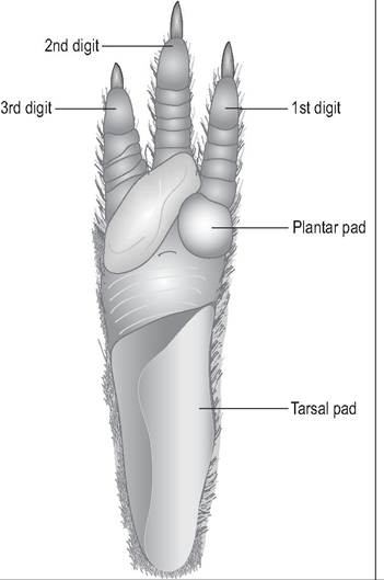
Figure 9.3 • Plantar surface of right hind foot of guinea pig.
with the sternum, ribs 10-13/14 being floating ribs (Breazile & Brown 1976; Cooper & Schiller 1975b)(Figs. 9.5 and 9.6).
Appendicular skeleton
The guinea pig has two tiny clavicles at the base of the neck and the acromion process of the scapula has an ‘L'-shaped hamate process. The bones of the forelimb are the humerus, radius and ulna, nine carpal bones, four metacarpal bones, and four digits.
The os coxa results from the fusion of four bones shortly after birth: the ilium, ischium, pubis, and acetabular bone. Each bone contributes to the formation of the acetabulum. There are sexual variations in the pubic symphysis. Immature males and females have a fibrocartilage articulation which becomes ossified over 1 year of age. In primiparous female guinea pigs the pubic symphysis remains cartilaginous so that it can dilate to allow the relatively large fetus to pass through. These pubic bones separate 2 weeks before parturition to allow the passage of the large fetus. Palpation of this can be a useful guideline for estimating time of parturition (Breazile & Brown 1976; Cooper & Schiller 1975b).
The hindlimb is composed of femur, tibia, fibula, eight tarsal bones, three metatarsals, and three digits that each have three phalanges. The patella is large. The thin fibula articulates proximally with the lateral condyle of the tibia and distally with the calcaneus.
Os penis
This is a small, thin rodlike bone that lies within the glans of the penis (Cooper & Schiller 1975b).
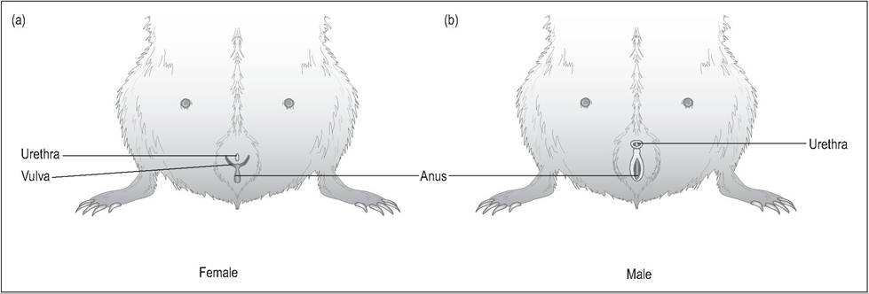
Figure 9.4 • External genitalia.
a) Female - There is a ‘Y’-shape with the vulva at the intersection of the branches and the anus distal to this. The urethral orifice lies between the branches of the Y.
b) Male - The cranial orifice is the penile urethra, covered by preputial folds. Caudally is a longitudinal cleft covering the opening of the large perineal sac and anus.
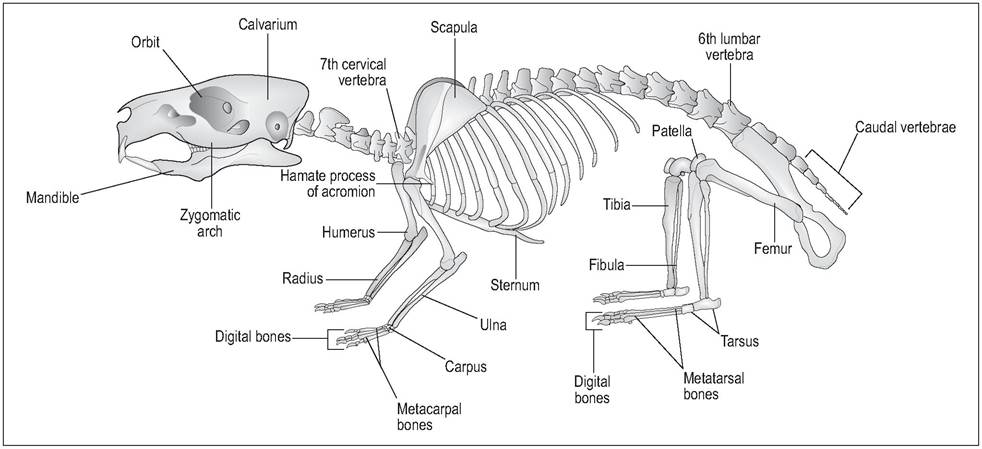
Figure 9.5 • Skeleton of guinea pig (Cavia porcellus) with major bones labeled. From Popesko, P., Rajtova, V., & Horak, J. (1990) A colour atlas of anatomy of small laboratory animals. Vol. I. Aylesbury, UK: Wolfe with permission.