CARDIOVASCULAR SYSTEM
The pericardium is thin and transparent, with fat deposits at the apex and insertion. It is firmly attached to the thymus cranioventrally. Due to the small size of the left lung the heart is exposed to the thoracic wall on this side (Bivin et al.
1979). There are two cranial vena cavae. The right vena cava empties directly into the right atrium while the left is joined by the azygos vein before joining the caudal vena cava to enter the right atrium (Bivin et al. 1979; Sharp & LaRegina 1998).The rat has the thinnest pulmonary artery and the thickest pulmonary vein of all rodent species examined. The vein is thicker due to cardiac striated muscle fibers that are continuous with those of the heart and which then extend into the lung tissue. This unfortunately could allow infectious agents to spread from the heart, through the pulmonary veins, and into the lungs (Bivin et al. 1979; Sharp & LaRegina 1998).
Venepuncture sites
The blood volume of the rat is 60 ml/kg and routine blood collection should be limited to 1% of body weight. Blood can be taken from the lateral tail vein, lateral saphenous or ventral tail artery. When intending to sample from the tail it should be warmed well first to cause vasodilation. As the
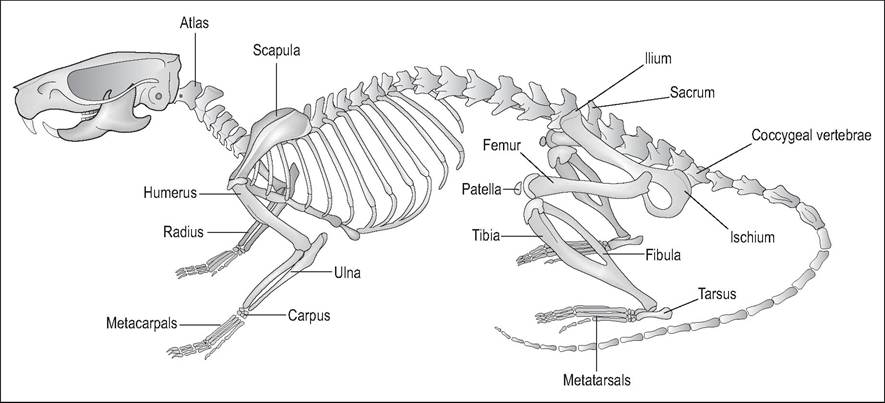
Figure 10.8 • Skeleton of Brown rat (Rattus norvegicus). From Popesko, P., Rajtova, V., & Horak, J. (1990) A colour atlas of anatomy of small laboratory animals. Vol. 2. Aylesbury, UK: Wolfe with permission.
ventral tail artery has high blood pressure no syringe plunger is necessary, although good hemostasis is required after removal of the needle to avoid excessive blood loss (Fallon 1996).