Cell Division
Mitosis
Each day millions of cells in the body of any normal animal die and are degraded or sloughed from epithelial surfaces. These cells must be replaced if normal life is to continue, and the replacement cells must be replicas of the original cells.
Mitosis, the division of somatic cells to produce two daughter cells, includes the duplication of genetic material for each daughter cell. Even though the process of cell division is normally a continuous process, here it has been divided into periods, or phases, for ease of communication about the process. The active phases are primarily based on nuclear changes visible by light microscopy. They are prophase, metaphase, anaphase, and telophase (Fig. 2-25).Interphase. The period between active cell divisions is the interphase. It may vary from a matter of hours in actively proliferating tissue to an almost permanent condition in cells that no longer divide, such as mature cardiac muscle cells. The replication of DNA during interphase prepares the cell to begin mitosis.
Prophase. Prophase, the first of the active phases, is characterized by condensation of chromatin into twisted filamentous threads (chromatids). (The term mitosis comes from the Greek word mitos, meaning thread.) Also during prophase, the nuclear envelope and the nucleolus begin to break down and disappear, and the two centrioles move to opposite poles of the cell. Microtubules become organized and arranged in a fan shape, radiating outward from the centrioles to the equator of the cell. This arrangement is the mitotic spindle.
Metaphase. Metaphase is the period when the nuclear envelope and nucleolus totally disap-
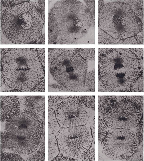
Figure 2-25. States of mitosis in cells of the whitefish blastula: 1) Interphase, with cell center adjacent to nucleus.
2) Early prophase, showing development of astral centers. 3) Late prophase, with astral centers at opposite ends of the cell. 4) Metaphase. 5) Early anaphase. 6) Late anaphase. 7) Early telophase. 8) Mid telophase, showing cleavage furrow. 9) Telophase interphase following separation of daughter cells. (Courtesy of Phillip G. Coleman, Michigan State University.)pear. The chromatids move and line up across the cell’s equator in the middle of the spindle, and the spindle microtubules attach to the centromere region of the chromatids.
Anaphase. Anaphase is the stage in which each centromere divides, separating the two chromatids, now properly called chromosomes again. The cell now contains twice as many chromosomes as it had originally. Half of the chromosomes begin to migrate toward one centriole at a pole of the spindle, and the other half migrates to the other centriole.
Telophase. Telophase begins when half of the chromosomes have been drawn by the microtubules to each pole of the cell. A nuclear envelope forms around each set of daughter chromosomes, and a nucleolus appears in each new nucleus. The spindle tubules disappear, and the chromosomes begin to unwind into filaments. Ultimately the chromosomes lose their visible identity and become the chromatin of the interphase period.
The cell itself next divides into two daughter cells. The division of the cytoplasm is called cytokinesis. It starts with invagination of the plasma membrane around the equator of the cell and ends by pinching off the two halves, with a nucleus in each half, creating the daughter cells. Each centriole is also replicated, and each daughter cell is now a replica of the parent cell. Mitosis is complete.
Meiosis
Meiosis (reduction division) differs from mitosis in a number of ways. it occurs during gametogenesis, the formation of ova in the female (oogenesis') and spermatozoa in the male (spermatogenesis). These processes are discussed in detail in Chapters 25 and 27. Since fertilization doubles the number of chromosomes in the fertilized ovum, equal numbers being contributed by the male and female, there must be a mechanism to reduce the somatic, or diploid, number of chromosomes in each gamete prior to fertilization.
Meiosis not only reduces the diploid number of chromosomes by half to the haploid number, it also increases the genetic variability of the offspring by crossing over. Homologous chromosomes in the primary sex (germ) cells pair up during prophase of meiosis. Homologous chromosomes are similar chromosomes that were contributed by the two parents of the individual. These paired homologous chromosomes may then cross over and exchange similar areas, resulting in two chromosomes that are different from either parent chromosome.