Nucleus
Structure of the Nucleus
The nucleus contains the genetic material of the cell encoded in molecules of DNA. With light microscopy, DNA and its associated proteins are seen as a more diffusely staining chromatin in the nondividing cell and as chromosomes in the dividing cell.
The nuclei of somatic cells contain the information necessary for determining the form and structure of new cells, and the nuclei of sex cells contain the information necessary to determine the characteristics of a new individual. The nucleoli consist largely of clustered DNA, which codes for ribosomal RNA; the nucleoli are seen as densely staining spherical bodies in the nucleus (Fig. 2-18).The nuclear envelope (Fig. 2-18), which surrounds the cell nucleus, is composed of two distinct membranes separated by about 20 nm. The outer membrane is continuous with the endoplasmic reticulum. Pores (small gaps or interruptions) in the nuclear envelope permit exchange between the protoplasm of the nucleus (nucleoplasm) and the cytoplasm outside the nucleus, including the movement of RNA synthesized in the nucleus out into the cytoplasm.
The functional activity and the continued life of the cell depend on the presence and functional integrity of a nucleus. A cell from which the nucleus has been removed (enucleated) gradually ceases activity, atrophies, and finally dies. However, if the nucleus is replaced with a nucleus from a cell from the same species prior to irreversible atrophy, function of the cell can be restored. The only cells in higher animals that do not have nuclei are mature red blood cells. This lack of nucleus is associated with their short life span, only 120 days.
The primary functions of the nucleus are (1) to regulate protein synthesis in the cell, thereby regulating the biochemical activities of the cell, and (2) to ensure the passage of genetic material (the chromosomes and their component genes) to subsequent generations of cells and/ or organisms.
DNA and DNA Replication
The genetic material necessary to direct cellular functions is primarily composed of chains of DNA. The chains of DNA are formed by joining small units (nucleotides), each containing a phosphate, a sugar (deoxyribose), and either a purine or pyrimidine base. The purine bases in DNA are adenine and guanine, and the pyrimidine bases are thymine and cytosine (Fig. 2-20).
Watson and Crick determined the structure of DNA to be a double helix, something like a spiral staircase or twisted metal ladder. The outside rails consist of two long chains of sugar-phosphate molecules, and the rungs are
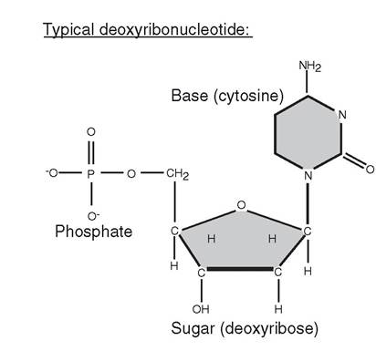
Figure 2-20. General structure of a nucleotide (subunits of DNA and RNA).
made up of paired bases that hold the two parts of the double helix together. Adenine is always paired with thymine, and guanine is always paired with cytosine. The two strands are joined by hydrogen bonds between the bases (Fig. 221). The two strands of the DNA double helix are not identical but are complementary. in other words, whenever adenine appears on one strand, thymine is in the same position on the opposite strand, and whenever guanine is on one strand, cytosine is in the same position on the other strand.
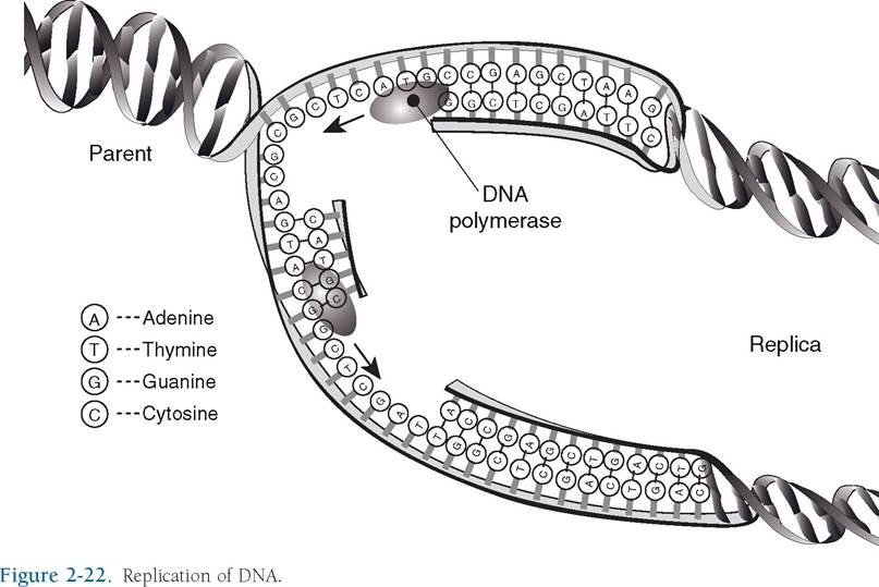
To pass on the genetic information to the next generation of cells or animals, the DNA double helix must be replicated. Replication of DNA begins with unwinding of the helix and splitting of the chain at the point of junction of
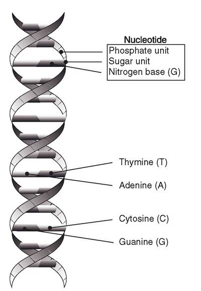
Figure 2-21. The basic structure of DNA. There are four nucleotides. The symbols in the upper right show how the nucleotides pair in DNA. (Adapted with permission from Cohen, B.J. and Wood, D.L. MemmlerS The Human Body in Health and Disease. 9th ed. Philadelphia: Lippincott Williams & Wilkins, 2000.)
complementary bases.
Each separated strand serves as a template, or model, for the formation of its complementary strand, which produces two double DNA helices, replicas of the original. Each new double helix consists of one strand of the original double helix and one newly synthesized strand (Fig. 2-22). Errors in the duplication of DNA strands during replication give rise to genetic mutations. Errors may occur spontaneously, or their frequency may be increased through the effects of numerous external factors, or mutagens (e.g., ionizing radiation, exposure to certain chemicals).The genetic information in DNA is coded by the specific sequence of purine and pyrimidine bases in a DNA molecule. This sequential arrangement of bases and its control of heredity, both on the cellular and the species level, have been called the genetic code, or the language of life. The interpretation of this code results in the synthesis of specific proteins. The only cellular constituent whose synthesis is specifically directed by the genetic code is protein. such proteins include those that are secreted as cellular products, those that are found in the cell membrane, and those that function within the cytosol or within cellular organelles.
The DNA code is said to be a triplet code, for each group of three nucleotides in the DNA chain ultimately calls for a specific amino acid in the process of protein synthesis. There are approximately 20 amino acids in the cell. With four bases that may be included in a triplet, there are more than enough potential triplet codes to represent the 20 amino acids. other triplet codes in the DNA serve as signals to demarcate the segment of the DNA chain that represents a particular protein and to regulate the initial and terminal steps in protein synthesis.
A gene is a segment of DNA that contains the triplet codes for all amino acids in one or more proteins and the signal sequences necessary to regulate the processing of the DNA segment. However, interspersed within a gene are also sequences of nucleotides that are not regulators of the process and do not contain necessary triplet codes.
These noncoding, non- regulatory segments are introns. The DNA coding segments in a gene are exons. A single gene may have multiple exons and introns throughout its length.RNA: Transcription and Translation
The processes by which the genetic code is interpreted and proteins are synthesized require the participation of three forms of RNA (ribonucleic acid). The three forms of RNA are messenger RNA (mRNA), transfer RNA (tRNA), and ribosomal RNA (rRNA). Like DNA, all three forms of RNA consist of nucleotide units that contain a sugar (ribose), a phosphate, and a purine or pyrimidine base. The two purines that are found in DNA, adenine and guanine, are also found in RNA, as is the pyrimidine cytosine. However, RNA does not contain the pyrimidine thymine that is found in DNA. Instead, RNA contains the pyrimidine uracil. Whereas the structure of DNA was two strands or chains of nucleotides joined together in a double helix, RNA exists only as a single strand.
The first step in the interpretation of the genetic code, transcription, results in the formation of a mRNA. This process is similar to DNA replication except that DNA now serves as a template for the synthesis of a mRNA instead of a new complementary strand of DNA. A specific nuclear enzyme, RNA polymerase II, and other nuclear proteins collectively known as general transcription factors bind together at a specific site on the DNA to initiate the transcription of a specific gene. The site on the DNA at which binding occurs and transcription begins is known as a promoter. A special segment of DNA adjacent to the promoter region contains a start sequence of nucleotides to signal RNA polymerase II to begin the synthesis of mRNA. Other regulatory proteins that act as enhancer or repressor transcription factors can also influence the rate of transcription of a specific gene. These regulatory transcription factors may bind to sites on DNA that are distant from the promoter region, but because of the folding and curling of the DNA strand, they can interact with the proteins bound at the promoter region.
Using the DNA as a template, RNA polymerase II synthesizes a single complementary strand of nucleotides. As with DNA, each group of three nucleotides in the newly synthesized mRNA is the code for a specific amino acid. Each group of three nucleotides in the mRNA is a codon. When the end of the segment of DNA that represents a particular protein is reached, a stop sequence in the DNA terminates the mRNA synthesis, and the mRNA strand detaches from the RNA polymerase. Figure 2-23 summarizes the steps of transcription.
Recall that the segment of DNA that represents a gene and serves as the template for mRNA contains both exons and introns (i.e., both coding and noncoding regions). Thus, the newly synthesized mRNA must be processed to remove the segments that correspond to the introns in the DNA. This processing is done before the mRNA exits the nucleus and enters the cytoplasm, where protein synthesis will take place. The segments of mRNA that correspond to the noncoding introns are excised, and the segments that correspond to the coding exons are spliced together by a complex consisting of RNA and a protein called a spliceo- some. During the splicing, the spliceosome sometimes omits segments of the initial mRNA that correspond to some of the coding exons. This omission results in alternately spliced mRNAs and ultimately the synthesis of different proteins from the transcription of a single gene.
Within the cytoplasm, the processed mRNA binds to a ribosome. This binding occurs at a specific end of the mRNA under the direction of a start codon at that site. The start codon also signals the binding of an initial tRNA with a specific amino acid attached. There is at least one specific tRNA for each of the approximately 20 amino acids in the cell. After the first tRNA is bound, a second tRNA with its attached amino acid arrives and binds to the next codon in line. Ribosomal enzymes then detach the amino acid from the first tRNA and link the two
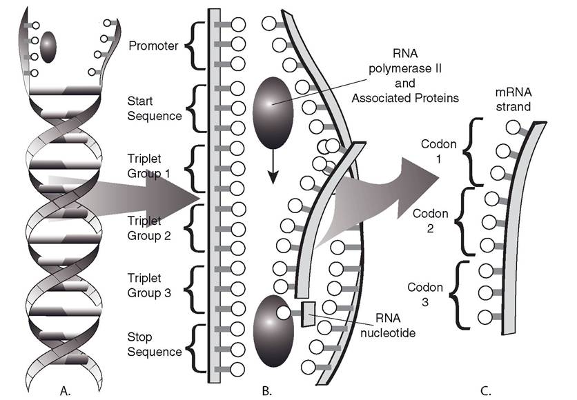
Figure 2-23.
Transcription. A) Separation of a DNA double helix. B) RNA polymerase II uses triplet groups as code to synthesize mRNA. C) Codons in completed mRNA correspond to triplet groups in DNA. A minimal number of triplets and codons are shown for clarity.amino acids to begin the formation of a peptide chain. The first tRNA can be detached from the ribosome and transfer another amino acid. The ribosome now directs the binding of a third tRNA with its appropriate amino acid and the subsequent linkage of the third amino acid to the second. Figure 2-24 summarizes these initial steps in protein synthesis.
This basic process of tRNA binding and amino acid linking continues as the ribosome moves along the mRNA strand. The result is a specific sequence of amino acids that are appropriate for the codons contained in the mRNA strand. A stop codon at the end of the mRNA signals the ribosome to detach the newly synthesized amino acid chain from the mRNA. The mRNA remains intact and may be reused multiple times. The decoding of the mRNA and the synthesis of the appropriate amino acid chain constitute translation.
Biotechnology
Genetic engineering and biotechnology are general terms used to describe the myriad of techniques used to alter the genetic code in organisms. Because the same principles of genetic information storage and transfer apply in all living organisms (from viruses through the hierarchy of plants, invertebrates, vertebrates, and humans), these techniques and procedures are widely applied. Because of the commonality of chemicals involved (DNA and RNA), it is also possible to move genetic material from one species to another. For example, mammalian DNA has been placed into the genome of Escherichia coli, which have then produced mammalian proteins. Recombinant DNA is the general term describing DNA that contains novel segments inserted by biotechnological techniques. An animal or
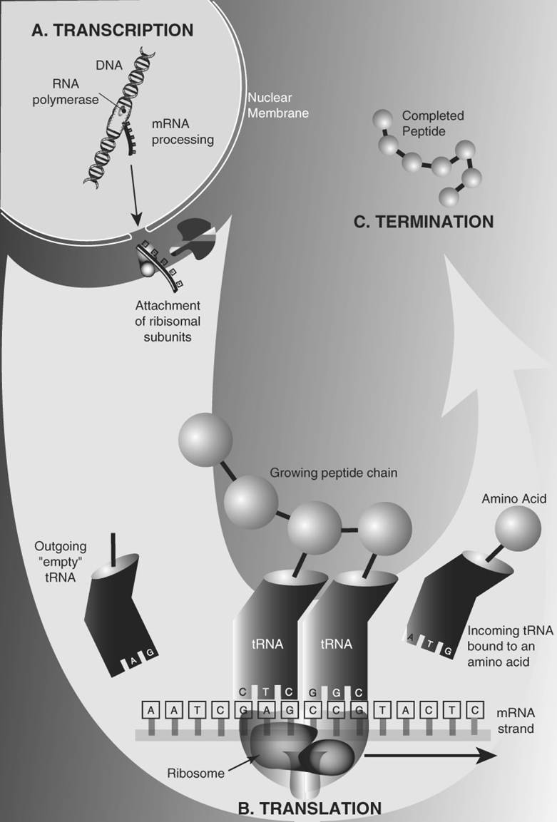
Figure 2-24. Protein synthesis. A) Transcription results in processed mRNA that exits nucleus. B) Translation of the mRNA produces a peptide chain. C) The completed peptide is released.
plant that contains DNA from another organism is said to be transgenic.
The discovery of a group of enzymes known as restriction nucleases was a key factor in the development of recombinant DNA techniques. These enzymes cut DNA into shorter segments by splitting the linkages between nucleotides. The enzymes do not act at random sites through the DNA strand; instead, each individual nuclease acts at a specific site termed its restriction site. If DNA from two different organisms are treated with the same nuclease, the nuclease will fragment the DNA at similar restriction sites in both. This yields segments of DNA with similar characteristics at their ends, but the sequences within the segments may be quite different. Because the ends of the segments from the two organisms are similar, DNA ligase (an enzyme that reestablishes the nucleotide linkages) can be used to join the different DNA segments. The final result is a DNA strand that contains DNA from two different organisms.
To produce a transgenic organism, recombinant DNA must be inserted into an organism’s genome. in domestic animals, this has been accomplished by microinjection of recombinant DNA into a pronucleus of single-cell embryos. The pronucleus is a nucleuslike structure in the embryo that contains genetic material from one parent. one-cell embryos have two pronuclei that ultimately fuse so that the genetic material from the two parents can be joined in the new individual. Recombinant DNA has also been transferred into embryos by infecting early-stage embryos with retroviruses containing recombinant DNA in their genome. Retroviruses are a specific group of viruses that insert their own genetic material into the genome of organisms they infect. As they insert their own genetic material, they also insert the recombinant DNA.
Clones are genetically identical individuals produced by asexual means. Cloning has been accomplished by splitting an early-stage multicell embryo into single cells, which continue their development into identical individuals. This technique has been successfully used in domestic animals. Clones have also been produced by nuclear transfer. In this technique, nuclei obtained from cells of adult animals are transferred into oocytes with their original nucleus removed. The oocytes with the transferred nucleus can be placed in the uterus of an appropriate female for gestation.