Cerebellar development in the neonatal animal
The ability of a neonatal animal to move correlates with the amount of cerebellar development at birth (Fig. 7.5). In precocious movers, such as herbivores that can ambulate within hours of being born, then cerebellar development is almost complete at birth.
It is essential that such animals can get up and run with the herd, thus they must be able to coordinate muscles used for posture and locomotion. In altricial animals, such as many predators, or burrow-born prey species (e.g. rabbits and rodents) cell division and migration are still occurring for several weeks after birth, and the cerebellum will not be fully functional for some time. This is reflected in their limited ability to move in a coordinated fashion in those first few weeks. Such animals are usually born into a protected environment such as a den or burrow.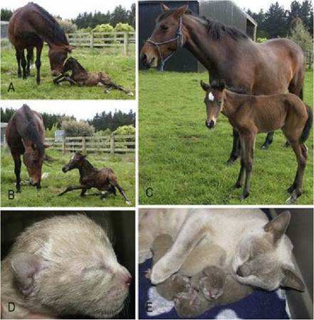
Fig. 7.5 The cat is an altricial animal, while the horse is precocial. The difference in mobility is largely related to the degree of cerebellar development at birth. Both the kittens and foal are less than 24 hours old.
(Cat photos courtesy of Ms. Genevieve Rogerson, Cahill Veterinary Clinic, Palmerston North. Horse photos courtesy of Dr. Debbie Prattley, IVABS, Massey University.)
Histology of the cerebellum
The cerebellum comprises four layers. Grey matter is found both superficially forming the cerebellar cortex, and in the three pairs of deep cerebellar nuclei.
The molecular layer is the most superficial. It is relatively acellular, being mainly cell processes, such as axons and terminations of afferent cells (e.g. granule cells, climbing fibres) and the dendrites of the efferent, Purkinje cells.
The Purkinje cells (also known as cerebellar piriform or pyramidal neurons) form the next layer. These cells have a large, pear-shaped cell body with a single axon projecting into the granule cell layer and multiple dendrites in the molecular layer.
The cell bodies form a line along the border between the molecular and granule cell layers. The cells are at a higher density at the tip of the folium than at the base of the folium. Purkinje cells are nestled in a layer of specialised astrocytes called Bergmann glia.The third layer is the granule cell layer comprising numerous, small-bodied neurons. It is a dense cell layer comprising 3-7 million granule cells/mm3. The thickness varies, being 5-6 cell layers at the bottom of the folium and 15-20 at the top of the folium.
The innermost fourth layer is the cerebellar white matter, in which are located the deep cerebellar nuclei, just above the fourth ventricle (Fig. 7.6 and 7.2, A21, A22).
Fig. 7.6
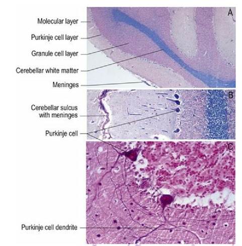
Histological sections of the cerebellum at different magnifications. (A, B, C) haemotoxylin and
eosin stain, Bielschowsky silver stain also on (C)
(from the Melvyn Birtle collection, IVABS, Massey University).
The functional histology of the cerebellum is complex, but in essence it comprises a system that receives afferent input from proprioceptors throughout the body and head, and also from the motor planning centres of the forebrain.
Afferent fibres to the cerebellum synapse on the deep cerebellar nuclei and excite them, or afferent input travels to the cerebellar cortex, where they stimulate the Purkinje cells or, they stimulate interneurons that inhibit Purkinje cells. Incoming fibres are descriptively named for their morphology and form mossy fibres (majority of fibres) or climbing fibres. Both mossy fibres and climbing fibres send collateral axons to the deep cerebellar nuclei as they ascend through the cerebellum to reach the cerebellar cortex. Mossy fibres are named for their mossy-like spreading appearance. They originate in brainstem nuclei and the spinal cord and ultimately synapse in the granule cell layer. The climbing fibres originate in the olivary nucleus of the medulla oblongata.
They decussate and send collaterals to the deep cerebellar nuclei, or climb all the way to the cerebellar cortex before synapsing.The output from the cerebellum is to motor planning centres in the forebrain, or to UMN centres of the motor cortex, or the UMN nuclei located throughout the brain. The output is largely from the deep cerebellar nuclei, which are facilitatory to the motor systems. However, Purkinje cells may inhibit the deep cerebellar nuclei, thereby blocking their facilitation of motor systems. The lateral and interposital nuclei project via the rostral cerebellar peduncle to the thalamus and red nucleus. The fastigial nucleus projects via the caudal cerebellar peduncle to the vestibular and reticular nuclei of the medulla oblongata.
The Purkinje cells form the only output from the cerebellar cortex. This output is primarily to the deep cerebellar nuclei. The exception to this is the output from the vestibulocerebellum (flocculonodular lobe) that projects directly to the vestibular nuclei of the brainstem, inhibiting their function. This arrangement is relevant to the pathogenesis of paradoxical vestibular disease (see Chapter 8). All Purkinje cells are inhibitory at their synaptic termination, mediated by the neurotransmitter GABA.
The functional histology of the cerebellum is summarised in Table 7.1 and Fig. 7.7. Overall, the cerebellum can function in two ways via the deep cerebellar nuclei. The nuclei may be stimulated by afferent cerebellar fibres or inhibited by the Purkinje cells. This results in the cerebellar nuclei being either facilitatory to motor systems or having no effect on them; they cannot inhibit them.
Table 7.1 The functional histology of the cerebellum
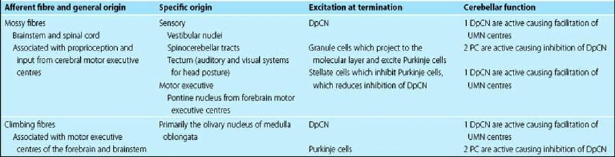
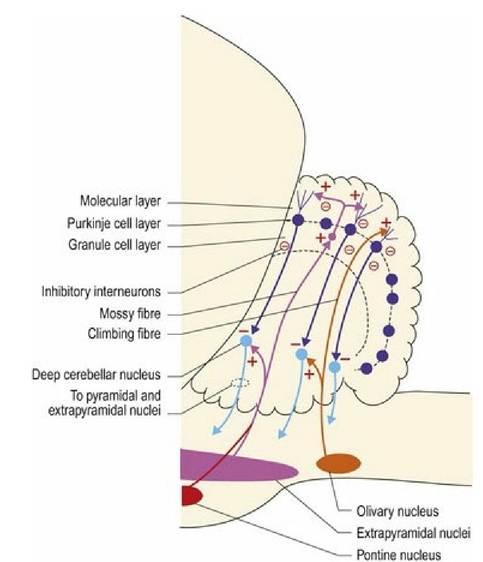
Fig. 7.7 Functional connections in the cerebellum. The red dots in the molecular and granule cell layers represent inhibitory interneurons that can inhibit Purkinje or granule cells. Note: for simplicity, only some connections with the brainstem are depicted.