CLASSIFICATION
1. What kinds of nerves are associated with the activity of smooth, cardiac, and skeletal muscle?
2. What is the principal distinguishing characteristic between smooth muscle and cardiac and skeletal muscle?
3.
What is the function of an intercalated disk in cardiac muscle?4. What is the functional difference between red and white skeletal muscle fibers?
There are three types of muscle cells of the animal body: smooth, cardiac, and skeletal. Each is characterized not only by microscopic structural differences, but also by location, function, and innervation.
Smooth Muscle
Smooth muscle is so named because it has no visible striations. The individual cells are spindle shaped and have a centrally located nucleus (Figure 8-1). Smooth muscles are regulated by the autonomic nervous system and are located in visceral structures that require movements of an automatic nature. Aggregates of myofilaments in smooth muscle are composed of the contractile proteins actin and myosin. The filaments are not arranged in order (as in skeletal muscle), which accounts for the lack of visible striations.
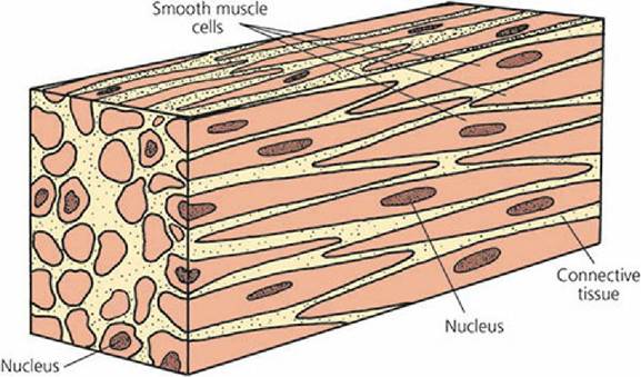
■ FIGURE 8-1 Smooth muscle cells exposed in their longitudinal and cross-sectional planes. The cells are characteristically spindle-shaped and have a centrally located nucleus.
Cardiac Muscle
Cardiac muscle is found only in the heart. It is regulated by the autonomic nervous system, like smooth muscle. In contrast to smooth muscle, however, on microscopic examination, cardiac muscle shows striations characterized by alternating light and dark bands. Cardiac muscle is composed of elongated, branching cells with irregular contours at their junctions with other cells, (Figure 8-2). The boundary area where the end of a cell anastomoses (joins) with the next cell is known as an intercalated disc.
This highly specialized cell membrane structure facilitates the transmission of nerve impulses from one cell to the next because of its low electrical resistance. Each cell has one nucleus (sometimes two) that is centrally located.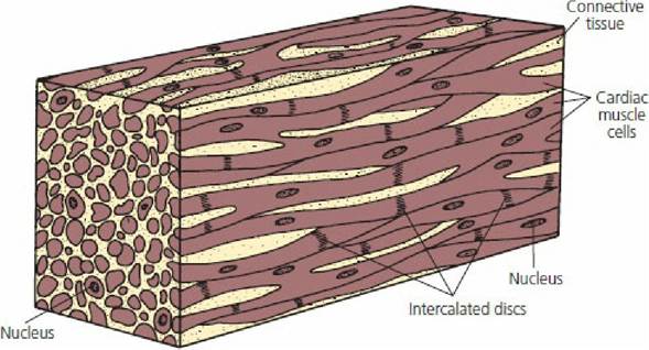
■ FIGURE 8-2 Cardiac muscle cells exposed in their longitudinal and cross-sectional planes. Note elongated, branching cells with irregular contours at their junctions with other cells, the intercalated disk. Intercalated disks indicate the positions of apposed borders of contiguous muscle cells. Most cells have a single nucleus, some contain two. The nucleus occupies a central position in the muscle cell.
Skeletal Muscle
Skeletal muscle cells (fibers) of many animals can be one of three types: (1) red or dark, (2) white or pale, or (3) intermediate, with characteristics between those of red and white fibers (Figure 8-3). Red muscle fibers are characterized by having more myoglobin and more mitochondria than white fibers. All muscles are probably a mixture of these three types, but in some animals the red and in others the white predominates. A striking example of this is the crimson red pectoralis muscle (breast muscle) of pigeons, which contrasts sharply with the stark white color of the chicken pectoralis muscle. Red muscle fibers usually contract more slowly and fatigue less readily than white muscle fibers. In birds, the amount of red pigmentation in the pectoralis muscle can be correlated directly with the ability to sustain flight. Geese and ducks, as well as pigeons, are known for their sustained flight and they have a predominance of red pectoralis muscle fibers.
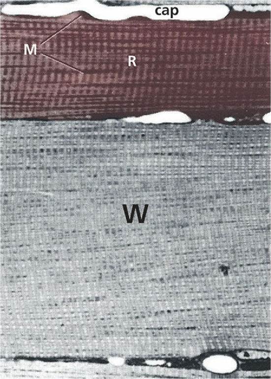
■ FIGURE 8-3 Photomicrograph of skeletal muscle showing red fibers (R) and white fibers (W). Red fibers have more mitochondria (M) packed between their myofibrils, especially in association with capillaries (cap).
(From Ham AW, Cormack DC. Histology. 8th edn. Philadelphia, PA: JB Lippincott, 1979.)Skeletal muscle makes up the major portion of the muscle mass of the animal body. An individual skeletal muscle fiber can extend the length of the muscle of which it is a part. As is characteristic of cardiac muscles, skeletal muscles are striated when viewed microscopically. They are not branched and do not anastomose (thus, no intercalated disk). Skeletal muscle is innervated by cranial and spinal nerves, and a nerve impulse to each muscle fiber is required for its stimulation. Multiple, peripherally arranged nuclei are present in each cell (Figure 8-4), in contrast to both smooth and cardiac muscle cells.
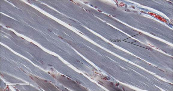
■ FIGURE 8-4 Photomicrograph of a longitudinal section of skeletal muscle fibers. Note the striations and the multiple, peripherally located nuclei.
■