COMPARISON OF CONTRACTION AMONG MUSCLE TYPES
1. What characteristic of muscle contraction is generally similar among smooth, cardiac, and skeletal muscle fibers?
2. Differentiate harnessing, innervation, and stimulus conduction between cardiac and skeletal muscle.
3. Do smooth muscle fibers have actin and myosin, a neuromuscular junction, a harnessing system, and a sarcotubular system?
4. How does the slower cycle of attachment and detachment of cross-bridge heads of smooth muscle relate to its function?
Brief structural differences among the three muscle classifications were noted earlier. The contraction process for all three is generally similar in that actin filaments slide between myosin filaments and cause a shortening of the cell. There is a greater similarity in arrangement of these filaments between cardiac and skeletal muscle (hence their common description as striated muscle). The myofibrils of cardiac muscle constitute most of the muscle fiber, but instead of being discrete and cylindric, as in skeletal muscle, they join together and are of variable diameter. This might be related to the more circular contraction of the heart (cardiac muscle) as compared with the more linear contraction of skeletal muscle.
Whereas the work of skeletal-muscle fibers is harnessed to connective-tissue elements, cardiacmuscle fibers anastomose with each other. Thus, the contraction of each joins with others to decrease the diameters of their respective heart chambers. Also, each skeletal-muscle fiber receives separate stimulation through a spinal or cranial nerve and neuromuscular junction, but cardiac muscle receives its stimulus from rhythmic, contractile, specialized cardiac-muscle cells known as pacemakers. The autonomic nervous system regulates the pacemakers. Conduction of stimulation is from cell to cell (through the intercalated disk) and special conduction fibers (Purkinje fibers in the ventricular walls).
The sarcotubular system of cardiac muscle is not as well developed as that of skeletal muscle.Smooth-muscle myofilaments are not aligned into myofibrils, as in cardiac and skeletal muscles. Furthermore, a higher ratio of actin to myosin exists (15:1 instead of 2:1). The actin filaments are attached to dense bodies that are dispersed inside the cell, and also some are attached to the cell membrane. The dense bodies correspond to the Z lines of skeletal muscle and are held in place by a framework of structural proteins that link one dense body to another. The actin filaments from two separate dense bodies extend toward each other and surround a myosin filament, thereby providing a contractile unit that is similar to a contractile unit of skeletal muscle (Figure 8-19).
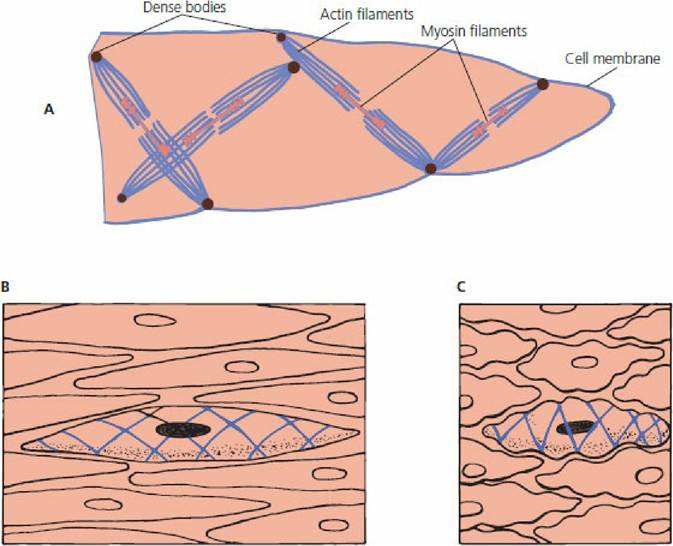
■ FIGURE 8-19 Contraction of smooth muscle. A. Physical structure of smooth muscle. Dense bodies attach either to the cell membrane or to an intracellular structural protein that links several dense bodies together. The dense bodies are functionally similar to Z lines. B. A translucent view of a relaxed smooth-muscle cell. C. A translucent view of a contracted smooth-muscle cell. Dense bodies and details of actin and myosin filaments not shown in B and C.
There are also differences between the contraction of smooth muscle and striated muscle. The cycle of attachment and detachment of cross-bridge heads that extend.Ifom myosin to actin is much slower in smooth muscle. This provides for prolonged tonic contraction, in contrast to rapid contractions of skeletal muscle. The slower cycles are a result of the much lower ATPase activity on the myosin cross-bridge heads than in skeletal muscle, and the heads remain in an “uncocked” position for a longer time. Coupled with the slower frequency of attachment-detachment cycling is the lower energy requirement for sustaining the same tension of contraction in smooth muscle as in skeletal muscle.
This is important from the standpoint of energy conservation, wherein smoothmuscle organs (e.g., urinary bladder, intestines) must maintain tone throughout the day and night.Smooth-muscle cells are able to shorten a much greater percentage of their total length than skeletal muscle. This feature enables a smooth-muscle organ, such as the urinary bladder, to reduce its lumen diameter from its expanded state to virtually zero.
The neuromuscular junctions associated with smooth muscle are diffuse junctions. The autonomic nerve fibers that innervate smooth muscle do not make direct contact with the muscle fibers but form diffuse junctions that secrete their transmitter substance into the interstitial fluid, whereupon it diffuses to the smooth-muscle cells. The vesicles of the terminal axons contain either ACh or norepinephrine, depending on whether the postganglionic terminal fiber is parasympathetic or sympathetic, respectively. The vesicle secretion may be excitatory or inhibitory depending on the receptors that are located on the smooth muscle membrane. The receptors determine whether the smooth muscle will be excited or inhibited and which one of the two transmitters will be effective in causing the excitatory or inhibitory response. The sarcotubular system of smooth muscle fibers is poorly developed. Counterparts to the T tubules are vesicles (called calveolae) that open on to the surface of the fiber just under the cell membrane.
■