SKELETAL-MUSCLE CONTRACTION
1. Describe the chain of events that initiates muscle-fiber membrane depolarization.
2. What is the association of Ca2+ to acetylcholine release? How is this related to milk fever in dairy cows?
3.
What is the trigger for release of Ca2+ from the sarcoplasmic reticulum? Where does Ca2+ go after release?4. Study the molecular basis of muscle contraction with emphasis on sequence and relate it to shortening and relaxation of muscle fibers.
5. What molecule seems to be necessary for relaxation?
6. What causes rigor mortis?
7. What is muscle tetany? Is this a reflection of motor unit summation or wave summation?
8. How is the bacterial disease, tetanus, related to central nervous system neurotransmitters?
Depolarization of Muscle Fibers
The neuromuscular junction functions as an amplifier for a nerve impulse. The arrival of a spinal or cranial nerve impulse at,the neuromuscular junction results in the release of acetylcholine (ACh) into the synaptic gap that is between the nerve-fiber terminal branch and the muscle fiber (see Figure 8-11). The release of ACh,is accelerated because calcium ions from the extracellular fluid enter the prejunctional membrane when the nerve impulse arrives. ACh is the stimulus that increases the permeability of the muscle-fiber membrane for sodium ions, after which membrane depolarization begins. Depolarization proceeds in all directions from the neuromuscular junction and an impulse is generated (see The Nerve Impulse and Its Transmission, Chapter 4). The impulse is conducted into all parts of the muscle fiber by the sarcotubular system (see previous text). Because the impulse initiates muscle contraction, a more synchronized contraction results when all parts of the fiber are depolarized nearly simultaneously as a result of sarcotubular transmission.
Almost immediately after its release, ACh is hydrolyzed by the enzyme acetylcholinesterase into acetic acid and choline. Acetylcholinesterase is highly concentrated in the synaptic cleft and this, coupled with the limited diffusion distance of ACh in the synaptic cleft, accounts for the rapid hydrolysis of ACh.
A low concentration of calcium in the extracellular fluid is recognized clinically in dairy cows after calving, known as parturient paresis, commonly called milk fever, as a state of semiparalysis caused by partial neuromuscular block. When the calcium ion concentration is low, the amount of ACh released is lowered; this might not be sufficient to cause neuromuscular transmission and thus neuromuscular block results.
Contraction Process
The contraction process involves an interaction between the actin filaments and myosin filaments. There is a natural attraction between actin and myosin that involves active sites on the actin filament. Attraction is inhibited during relaxation because the active sites are covered, but when calcium ions enter the myofibril, the active sites are uncovered. The projecting portions of the myosin filament cross-bridge heads attach to the active actin filament sites during contraction and bend toward the center, causing actin to slide toward the myosin filament center. The relative location of the actin and myosin myofilaments to each other within a sarcomere is shown in Figure 8-13.
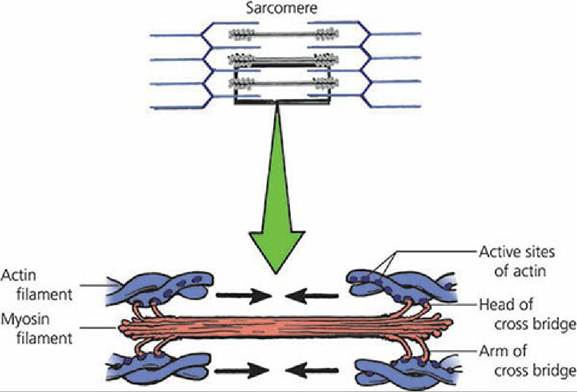
■ FIGURE 8-13 The components of the actin and myosin myofilaments associated with contraction of the sarcomere. Arrows indicate the direction of actin movement during contraction (shortening of myofibrils).
The actin filament has three major components (all protein): actin, tropomyosin, and troponin (Figure 8-14A). Actin and tropomyosin are arranged in helical strands interwoven with each other. Troponin is located at regular intervals along the strands and contains three proteins, two of which bind actin and tropomyosin together and the third that has an affinity for calcium ions.
Active sites (places where myosin cross-bridges attach) are located on the actin strands and are normally covered by the tropomyosin strands (Figure 8-14B). When calcium ions bind to the troponin complex, a conformational change occurs between the actin and tropomyosin strands and causes the active sites to be uncovered. The uncovered sites favor activation of the natural attraction that exists between actin and myosin and allows attachment of myosin cross-bridge heads (Figure 8-14C). The interaction of the actin filaments and the myosin filaments showing a cycle of contraction followed by relaxation are shown in Figure 8-15.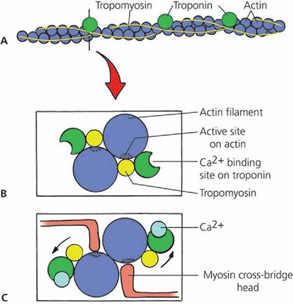
■ FIGURE 8-14 Conformational changes of the actin filament after calcium binding. A. The actin filament with its three proteins, actin, troponin, and tropomyosin. The vertical line indicates the cross-section location for B and C. B. The active sites on actin are covered by tropomyosin. C. Ca2+ binds to troponin, resulting in a conformational change that exposes the active sites on actin. Myosin cross-bridge heads attach to actin active sites and myofibril contraction begins.
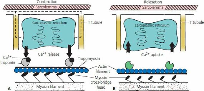
■ FIGURE 8-15 A cycle of contraction followed by relaxation. A. The dashed line indicates transfer of depolarization from the sarcolemma and T tubules to the sarcoplasmic reticulum. Depolarization is followed by Ca2+ release from the sarcoplasmic reticulum with diffusion to the myofibrils. Ca2+ binds to troponin, removing the blocking action of tropomyosin. Myosin crossbridge heads attach to active sites on actin and bend toward the center of the myosin molecule. B. Relaxation begins when ATP binds to cross-bridge heads, causing their detachment from actin. Ca2+ is returned to the sarcoplasmic reticulum using energy supplied by ATP.
Removal of Ca2+ from troponin restores the blocking action of tropomyosin.Energy Changes
The energy changes that permit attachment and detachment of the myosin cross-bridge heads are synchronized with the mechanical changes of the actin molecule during contraction and relaxation. These are summarized as follows and illustrated in Figure 8-16 (assume that cross-bridge heads have just bound with adenosine triphosphate (ATP) and have detached from the active sites of the actin filaments):
1. Adenosine triphosphatase (ATPase) of the myosin cross-bridge heads hydrolyzes ATP to adenosine diphosphate (ADP) + inorganic phosphorus (Pi), leaving the ADP + Pi bound to the heads. Energy from the hydrolysis of ATP “cocks” the heads so that they increase their angle of attachment to the cross-bridge arm and become perpendicular to the active sites of the actin myofilaments (Figure 8-16A).
2. After depolarization of the sarcotubular system, calcium ions diffuse from the sarcoplasmic reticulum into myofibrils and bind to the troponin, and the actin filament active sites are uncovered, removing the blocking action of tropomyosin. The natural attraction of myosin to actin is now permitted and the “cocked” heads bind with active sites (Figure 8-16B).
3. Binding with actin causes a conformational change in the heads (“uncocking”), and they bend (tilt) toward the cross-bridge arms (toward the center of the sarcomere), pulling actin with them. Energy for this is derived from previous ATP hydrolysis. Calcium ions are returned rapidly to the sarcoplasmic reticulum once the shortening process begins (Figure 8-16C).
4. Tilting of the cross-bridge heads causes release of ADP and Pi and sites on the heads are exposed for binding of new ATP. The binding of new ATP causes detachment of myosin cross-bridge heads from actin myofilaments (Figure 8-16D).
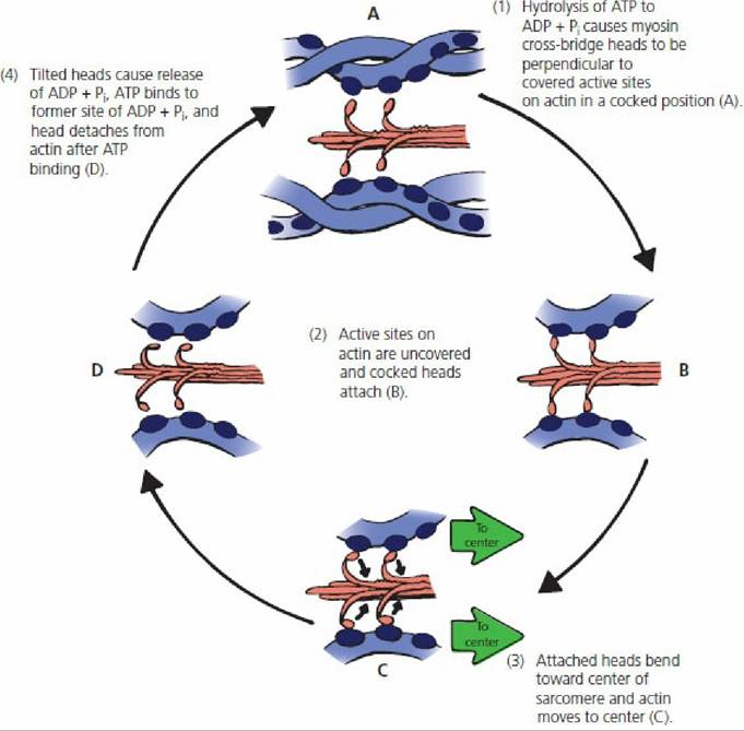
■ FIGURE 8-16 Energy changes associated with actin and myosin interaction that result in muscle shortening.
See text for details. ATP, adenosine triphosphate; ADP, adenosine diphosphate; Pi, inorganic phosphorus.The ATPase of myosin cross-bridge heads then hydrolyzes ATP as before, cocking the heads; the process is repeated when the next neuromuscular transmission causes depolarization of the sarcotubular system. Repetition of the process causes the actin myofilaments to be pulled farther into the center, thus shortening the sarcomere.
The immediate energy for muscle contraction is thus derived from ATP, forming ADP + Pi. The amount of ATP in muscle fibers is limited and rephosphorylation of ADP must occur so that contraction can continue. This is accomplished by transfer from creatine phosphate (CP), which is about five times more plentiful than ATP, according to the following reaction:
CP÷ADP bn*t >C÷ATP
Because the amount of CP is also limited, the necessary rephosphorylation of creatine (C) and ADP is ultimately derived from intermediary metabolism within the muscle cell and from the associated reoxidation of reduced cofactors that occurs in the electron transfer chain of the mitochondria. The presence of ATP is required for relaxation, or detachment of the myosin from the actin, and also for the return of calcium ions to the sarcoplasmic reticulum.
Muscle contraction is 50% to 70% efficient in regard to the accomplishment of work. The nonwork portion is dissipated as heat. This heat source is important to the body for the maintenance of body heat. Body cooling results in shivering, which is an attempt by the body to generate heat by muscle contraction.
Contraction versus Contracture
Muscle shortening can occur in the absence of action potentials. This type of shortening is referred to as rigor or physiologic contracture, as opposed to contraction. The actin and myosin filaments remain in a continuous contracted state because sufficient ATP is not available to bring about relaxation (see previous text). Contracture that occurs after death is referred to as rigor mortis.
Lack of ATP for relaxation in this case endures, however, and relaxation only occurs as a result of postmortem autolysis caused by enzymes released from the lysosomes 12 to 24 hours after death. Those muscles that were most active just before death are those that develop rigor mortis first (i.e., greater exhaustion of ATP and CP associated with greater muscle activity).Contraction Strength
Contraction strength varies and is achieved by motor unit summation or by wave summation. The stimulation of one motor unit causes a weak contraction, whereas the stimulation of a large number of motor units develops a strong contraction. This is known as motor unit summation. All gradations of contraction strength are possible, depending on the number of motor units stimulated. Increasing the strength of contraction by wave summation occurs when the frequency of contraction is increased. When a muscle is stimulated to contract before the muscle has relaxed, the strength of the subsequent contraction, as measured by the height of a lifted load, is increased. When the frequency is sufficient such that the individual muscle twitches become fused into a single prolonged contraction, the strength is at a maximum; this condition is known as tetany (Figure 8-17).
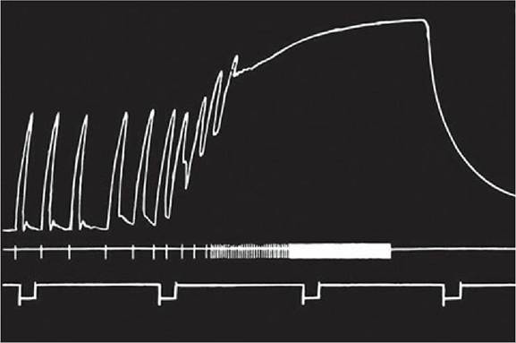
■ FIGURE 8-17 Increasing muscle strength by increasing the frequency of contraction. This is known as wave summation. Tetany occurs when individual contractions are fused and cannot be distinguished from each other. (From Carlson AJ, Johnson V. The Machinery of the Body. 4th edn. Chicago, IL: University of Chicago Press, 1953.)
Tetanus
Tetanus is a bacterial disease caused by a potent neurotoxin elaborated by the organism Clostridium tetani. The neurotoxin reaches the central nervous system and prevents release of an inhibitory transmitter (glycine). The resulting sensitivity to excitatory impulses, unchecked by inhibitory impulses, produces generalized muscular spasms (tetany). Tetanus has been called lockjaw because the masseter muscles that close the mouth are stronger than the muscles that open the mouth and the jaws remain in a closed (locked) position.
Treppe
Muscles seem to “warm up” to a maximum contraction state. This can be shown by applying stimuli of equal intensity a few seconds apart to a muscle. Each successive muscle twitch has slightly more strength than the previous one, until optimal contraction strength is reached (Figure 8-18). This phenomenon is referred to as treppe, or the staircase phenomenon. Successive stimulations are believed to provide for an increasing concentration of calcium ions in the sarcoplasm during the beginning contractions of rested muscles.
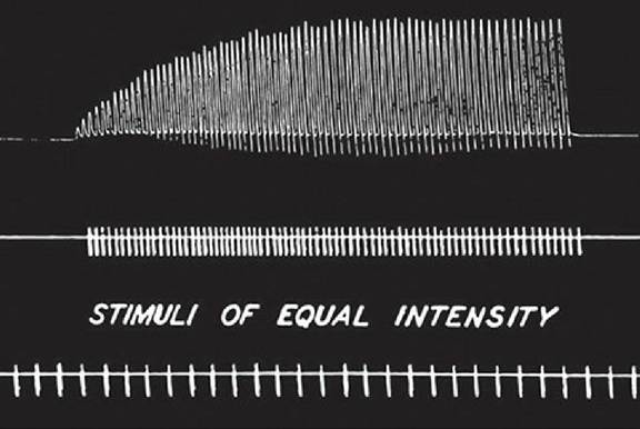
■ FIGURE 8-18 The staircase phenomenon of skeletal muscle. This is also known as treppe. Successive stimuli of the same intensity produce contractions of increasing strength. (From Carlson AJ, Johnson V. The Machinery of the Body. 4th edn. Chicago, IL: University of Chicago Press, 1953.)