» Conformation and Superficial Features
The head and neck together form a cone that blends with the trunk at the level of the forelimbs. The skull of primitive breeds, as of the ancestral wild form, is more or less pyramidal, but that of most improved breeds sweeps sharply upward to a prominence that rises well above the brain (Fig.
32.1). The dorsal surface of the cranium is bounded caudally by a thick nuchal crest and demarcated from the temporal fossa to each side by a prominent temporal line that continues into the zygomatic process of the frontal bone. This process, relatively short, fails to meet the zygomatic arch, which completes the margin of the small orbit (see Fig. 32.8). The arch is extremely sturdy and carries the wide, flat articular surface and, more rostrally, the depression from which the levator labii superioris arises.On the basal surface, the cranial and choanal regions of the skull are dorsal to the plane of the palate. The large paracondylar processes and tympanic bullae are prominent features of the cranium. The body of the stout, rather rectilinear mandible is cut away in adaptation to the rooting habit. The mandibular symphysis ossifies at about 1 year.
The most striking feature of the head is the rostrum, or snout, the disklike and mobile tip of the muzzle that incorporates the middle part of the upper lip and is perforated by the rounded nostrils (Fig. 32.2). The snout is supported by a small rostral bone set against the end of the nasal septum that gives attachment to the levator labii superioris (Fig. 32.3/3), the muscle principally concerned with movements of the snout. Pigs allowed access to open ground are generally "ringed" through the upper margin of the snout to discourage the rooting habit, a practice more frequently required in former times than today. The lips are short and rather immobile; the upper one is notched to accommodate the projecting canine tooth (tusk).
The small eyes are deeply placed and, uniquely among domestic species, lack a tapetum lucidum and therefore are not reflective of light. A deep lacrimal gland is associated with the third eyelid in the ventromedial angle of the orbit. Together with the retrobulbar muscles, it is engulfed by an orbital venous sinus that may be punctured at the medial angle of the eye by directing a needle medioventrally, between the globe and the third eyelid. The procedure is most likely to be performed in a research context. The sinus is said to be involved in thermoregulation of brain temperature by conveying cool blood from the nasal cavity.
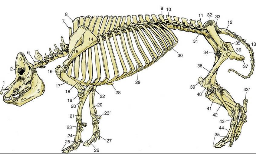
FIG. 32.1 Skeleton of a pig. 1, Rostral bone; 2, orbit; 3, temporal fossa; 4, zygomatic arch; 5, mandible; 6, first cervical vertebra; 7, last cervical vertebra (C7); 8, first thoracic vertebra; 9, last thoracic vertebra (T16); 10, first lumbar vertebra; 11, last lumbar vertebra (L5); 12, sacrum; 13, caudal vertebrae; 14, scapula; 15, spine of scapula; 16, greater tubercle of humerus; 17, humerus; 18, sternum; 19, condyle of humerus; 20, radius; 21, ulna; 22, olecranon; 23, carpal bones; 23', accessory carpal bone; 24,
metacarpal bones; 25, phalanges; 26, phalanges of principal digit; 27, phalanges of accessory digit; 28, xiphoid cartilage; 29, 10th pair of ribs; 30, costal arch; 31, coxal tuber; 32, iliac crest; 33, sacral tuber; 34, head of femur in acetabulum; 35, ischial spine; 36, greater trochanter; 37, ischial tuber; 38, femur; 39, patella; 40, lateral condyle of femur; 41, tibia; 42, fibula; 43, tarsal bones; 43', calcaneus; 44, metatarsal bones.
The oval ears are attached to the high caudal part of the head and in lop-eared breeds hang down over the face. The external surface displays the only veins convenient for intravenous injection. These may be readily visible but, if not, are made so by application of a tourniquet at the base of the ear.
The lateral vein of the set is most often used. Chewing of their companions' ears is a common vice among young pigs raised together in close quarters.Subcutaneous injections are commonly made at a site just caudal to the ear; awareness of the proximity of the parotid gland is necessary (Fig. 32.3/15). The same site is used for injection into the muscle mass directly caudal to the skull; however, the orientation of the needle is different.
The neck is roughly cylindrical but with some lateral compression. It is remarkably short; the closeness of the angle of the mandible to the shoulder joint prevents the animal from turning its head to any great degree. The flabby lateroventral parts of the neck, the jowls, are common seats of abscesses.
The more important superficial structures of the head are shown in Fig. 32.3. They include the buccal branches of the facial nerve (Fig. 32.3/19 and 20); the ventral one follows a course around the lower margin of the masseter in company with the parotid duct and the facial artery and vein. The artery is short because the dorsal part of the face is supplied by the infraorbital artery that reaches the region through the infraorbital foramen together with the nerve of the same name. The facial vein is partly formed by a frontal tributary that becomes superficial by emerging through the foramen dorsomedial to the orbit. As would be expected, the infraorbital nerve is large because it supplies the sensitive snout.
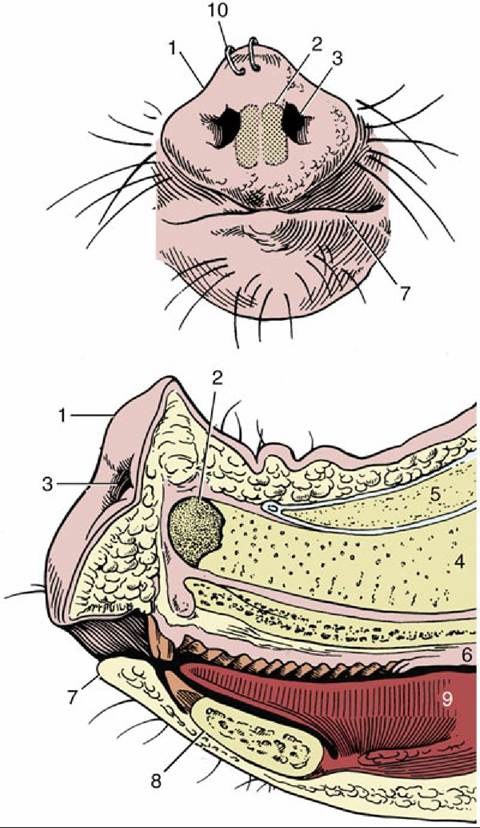
FIG. 32.2 The snout from the front and in median section. 1, Rostral plate; 2, rostral bone; 3, nostril; 4, nasal septum; 5, nasal bone; 6, hard palate; 7, lower lip; 8, mandible; 9, tongue; 10, nose rings to discourage rooting.