Corpus Luteum
The ovarian corpus luteum (pl. corpora lutea) is a temporary endocrine organ with progesterone as its primary secretory product. A corpus luteum forms at the site of each ovulated follicle (Fig.
27-1), so litter-bearing animals may have multiple corpora lutea on an individual ovary.sometimes during ovulation small blood vessels rupture, and the cavity of the ruptured follicle fills with a blood clot, a corpus hemor- rhagicum. Whether or not a corpus hemor- rhagicum forms, the granulosa cells lining the empty follicular cavity begin to multiply under the influence of LH and form a corpus luteum, or yellow body. The granulosa cells also continue to undergo luteinization. Most luteal cells are derived from granulosa cells, but some cells in the corpus luteum are derived from the theca interna.
Although a mature follicle and a fully formed corpus luteum are about the same size, they can be differentiated by sight or palpation. The follicle is a sac filled with fluid that has the appearance and feel of a blister, while the corpus luteum looks and feels solid (Fig. 27-5).
Blood progesterone levels increase as corpora lutea grow and develop after ovulation (Fig. 27-2). When corpora lutea are fully developed, progesterone secretion is maximal and plasma levels stabilize. If fertilization of the ova does not occur and a pregnancy is not established, the corpora lutea spontaneously regress, with a relatively rapid decrease in plasma progesterone (Fig. 27-2). Corpus luteum regression entails apoptotic death of luteal cells, their removal, and the replacement of the corpus luteum with connective tissue forming a corpus albicans. If a pregnancy is established, maternal recognition of pregnancy occurs, and regression of the corpus luteum is prevented. This process and the role of the corpus luteum during pregnancy are discussed in more detail in Chapter 28.
The basic function of progesterone during this part of the estrous cycle is to prepare for a pregnancy. Progesterone increases uterine gland secretion and inhibits uterine motility to promote implantation and maintain any pregnancy (Fig. 27-2). Progesterone also promotes mammary gland development. High levels of progesterone act on the hypothalamic- adenohypophyseal axis to inhibit further LH secretion.
If a successful pregnancy is not established, the corpora lutea must undergo regression
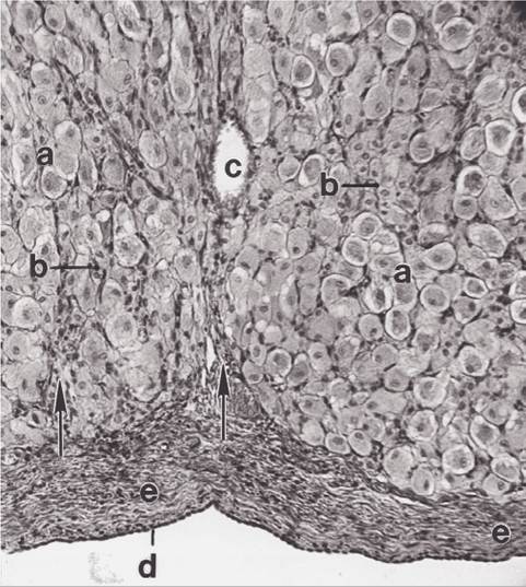
Figure 27-5. Part of a mature corpus luteum (sow). Two types of cells, large luteal cells (a) and small luteal cells (b) can be identified. A blood vessel (c) enters from the periphery and trabeculae (arrows). The ovarian epithelium (d) and adjacent tunica albuginea (e) are also shown. (Reprinted with permission of Wiley-Blackwell from Dellmann H.D. and Eurell J. Textbook of Veterinary Histology. 5th ed. Baltimore: Lippincott Williams & Wilkins, 1998.)
(luteolysis) to permit the animal to continue the estrous cycle. The humoral signals between the uterus and ovary that initiate or inhibit luteoly- sis differ among species. In most domestic species (mare, cow, ewe, sow), prostaglandin F2a (PGF2a) is the humoral signal used by the nonpregnant uterus to stimulate luteolysis. The nonpregnant uterus increases PGF 2α synthesis; releases are increased after ovulation at times appropriate for the species (e.g., 10 days for sows and 14 days for ewes); and luteolysis occurs shortly thereafter (Fig. 27-2).
Luteolysis can be induced in cattle by administering analogs of PGF2a at any point in the estrous cycle as long as a corpus luteum is intact and functioning. The removal of the corpus luteum permits rapid development of new follicles and ovulation in about 3 days. The use of PGF2a to induce ovulation and estrus at a predictable time is a management tool to synchronize the estrus cycles of groups of animals.