Ovulation
In mature follicles just prior to ovulation, ova are usually seen surrounded by a halo of granulosa cells (cumulus) that are continuous with granulosa cells lining the fluid-filled antrum (Fig.
27-3). The large, thin-walled follicles bulge from the ovarian surface (Fig. 27-4).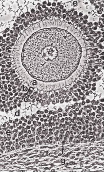
Figure 27-3. Bovine oocyte shortly before ovulation. Oocyte is surrounded by zona pellucida (a), the corona radiata (b), a single layer of unique granulosa cells immediately adjacent to the zona, and a layer of granulose cells termed the cumulus oophorus (c). The cumulus appears to have separated from the granulosa cells lining the antrum of the follicle (e). The theca interna (g) is also visible. (Reprinted with permission of Wiley-Blackwell from Dellmann H.D. and Eurell J. Textbook of Veterinary Histology. 5th ed. Baltimore: Lippincott Williams & Wilkins, 1998.)
The primary oocyte, which remains in an arrested stage of meiosis during follicular development, undergoes the first meiotic division to produce a secondary oocyte and the first polar body just before ovulation in most species. The first polar body is extruded from the ovary with the secondary oocyte. In the mare, this first meiotic division occurs just after ovulation.
Luteinizing Hormone Surge
In most species, LH release from the adenohypophysis increases sevenfold to tenfold during the 24 hours prior to ovulation. After reaching its peak, LH release rapidly decreases and plasma levels return to preovulatory levels (Fig. 27-2). This short-term change in LH release is the LH surge. As discussed earlier, the LH surge depends on changes in the hypophyseal- adenohypophysis axis and an increase in adenohypophysis content of LH induced by the rapid rise in production of estrogens by large, mature follicles.
The extremely high levels of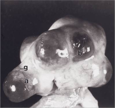
Figure 27-4. Ovary of sow with mature tertiary follicles just before ovulation (a) and a site where a follicle has just ovulated (b). A surface vessel is also visible (g). (Reprinted with permission of Wiley-Blackwell from Dellmann H.D. and Eurell J. Textbook of Veterinary Histology. 5th ed. Baltimore: Lippincott Williams & Wilkins, 1998.)
LH promote the final development of the primary oocyte and its progress though the first meiotic division. This prepares the oocyte for ovulation.
Granulosa cells also respond to the LH surge by transforming from estrogen-producing cells to progesterone-producing cells. This is part of luteinization, the transformation of granulosa cells to luteal cells (cells of a corpus luteum). This process begins prior to ovulation, so estrogen levels are decreasing and progesterone levels are increasing at ovulation. Under the influence of the LH surge, granulosa cells also acquire the ability to synthesize prostaglandins, thromboxanes, and leukotrienes. These agents induce a local response similar to inflammation that will weaken the wall of the follicle and promote its rupture.
Spontaneous and Reflex Ovulators
The LH surge and ovulation occur in most domestic species (mare, cow, ewe, and sow) independent of copulation, and these species are spontaneous ovulators. In these species, the preovulatory increase in estrogens from developing follicles is the primary event that brings about ovulation. The female animals of some species (rabbit, ferret, mink, camel, llama, and alpaca) usually require copulation for ovulation. These are induced ovulators. In these species, the final preovulatory surge of GnRH, and subsequent LH surge, is apparently dependent on a neural reflex elicited by vaginal stimulation. Induced ovulators have characteristic estrous cycles and follicular development, but mature follicles regress if copulation does not occur.