Development of Germ Layers
The one-celled zygote undergoes the first mitotic divisions, known as cleavage, shortly after fertilization. Cleavage increases the number of cells (called blastomeres) without increasing the volume of the developing embryo, so that after each cell division, the daughter cells have smaller cytoplasmic mass.
However, the nuclei of the daughter cells are normal in size and contain a full complement of chromosomes. The cluster of small cells resulting from cleavage has a lobulated appearance resembling a berry; hence, the name morula (Latin, small mulberry) is given to this stage (Fig. 3-1).When the morula reaches the uterus, a cavity, the blastocele, forms within it, transforming the morula into a hollow ball called a blastula. The blastula comprises a layer of cells, the trophoblast, surrounding the blastocele, into which a collection of cells, the inner cell mass, protrudes. The inner cell mass eventually forms the body of the embryo. The trophoblast will develop into the extraembryonic tissues, including the placenta.
Because they retain the potential to become any cell of the embryo (excluding the extraem- bryonic tissues), the cells of the inner cell mass are often described as being pluripotent. it is these cells of the early embryo, the so-called embryonic stem cells, that are the subject of such intense interest and debate in the scientific community (see p. 57).
It is the blastula stage that is collected from donor animals for embryo transfer.
The portion of the inner cell mass closest to the trophoblast is the epiblast, and the portion adjacent to the blastocele is the hypoblast. As the inner cell mass develops, a cavity forms dorsal to the epiblast; this is the amniotic cavity.
sometime before the end of the second week of development, the epiblast begins to thicken
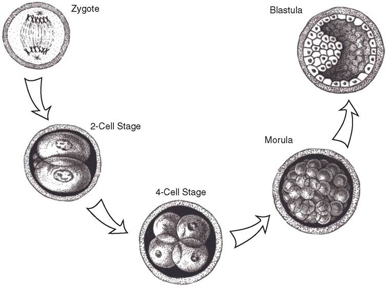
Figure 3-1. Development from zygote to blastula.
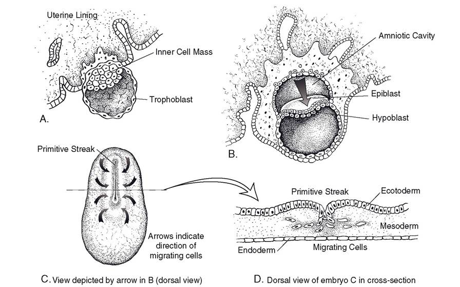
Figure 3-2. Gastrulation. A) and B) Around the time of implantation, when the embryo embeds in the wall of the uterus, the inner cell mass becomes a disk of two distinct layers, epiblast and hypoblast. C) Embryo viewed from above, as indicated by the arrow in B. Cells of the epiblast begin to proliferate and migrate toward the longitudinal primitive streak on the dorsal midline. D) Cross-section through the region of the primitive streak. Migrating cells move to the interior of the embryo, where they become mesoderm.
with proliferating cells on the longitudinal axis of the embryo. This thickening is the primitive streak, and here the epiblast cells migrate into the interior of the embryo, taking up residence deep to the outer layer of cells (now called ectoderm) and displacing the hypoblast to create a deep layer, the endoderm. Between the ectoderm and endoderm the third and final germ cell layer, the mesoderm, is established. This migration of cells is gastrulation (Fig. 32); with it the embryo establishes the three primary cell lines that will give rise to all of the tissues in the adult body.
The ectoderm on the dorsal surface of the embryo will become epidermis and nervous tissue. The endoderm is the germ cell line destined to become the lining of the gastrointestinal and respiratory systems, the epithelial parts of glands associated with the digestive system, and parts of the reproductive system. The mesoderm gives rise to muscle, the skeleton, urinary and cardiovascular systems, and parts of the reproductive system.