DEVELOPMENT OF THE URINARY ORGANS
The intermediate mesoderm reflects in muted fashion the segmentation that is so evident in the adjoining somites. It soon forms in its caudal domain a continuous solid longitudinal (nephrogenic) thickening from which arise, in craniocaudal and temporal sequence, three attempts at the formation of an excretory organ.
The first attempt constitutes the pronephros, which forms in the presumptive neck region; this has a transient existence and is not functional in mammals. The second attempt, the mesonephros, forms in the thoracic and lumbar regions and is more successful; it is functional through a large part of embryonic life. The third attempt, the metanephros, forms in the lumbar region; it becomes the adult kidney (Figure 5-3).All three structures have a series of excretory tubules as their essential histological feature. In the pronephros one end of each tubule turns caudally to meet its neighbor, and in this way a continuous pronephric duct is formed (Figure 5-3/4), which at its caudal end grows toward and opens into the cloaca. The duct survives the regression of the pronephric tubules and is adopted as the means of drainage of the mesonephric tubules that now appear. Because the pronephric tubules are nonfunctional, their peculiarities of construction need not be noted.
The mesonephric tubules are much more numerous. Each resembles a rather simple version of the nephron of the adult kidney in structure and function (see Figure 5-27). The blind end is invaginated by a capillary tuft to form a filtration mechanism while the connection of the other end with the pronephric duct, now more appropriately termed the mesonephric duct, provides an outlet for the urine that is formed. The mesonephros may be a very prominent organ at its apogee, when it projects from the roof of the abdomen (Figure 5-4). Its size varies among species and is in inverse proportion to the permeability (and thus the excretory efficiency) of the placenta.
The mesonephros is supplanted by the metanephros when it begins to regress, which is a process that occurs in a craniocaudal direction. Parts, however, survive to be given fresh use by the male reproductive system (Figure 5-5).The metanephros has two primordia. One is provided by an outgrowth, the ureteric bud, from the lower end of the mesonephric duct close to its opening into the cloaca. This bud grows cranially into the metanephric blastema constituted by the caudal part of the nephrogenic cord (Figure 5-3/5). The extremity of the bud undergoes a dozen or so dichotomous divisions. Branches of the later orders become the collecting tubules of the kidney, whereas those of the first few orders are later reabsorbed into the terminal expansion of the duct in a variable fashion that accounts for the specific forms of the renal pelvis and calices. The outer part of the metanephric mass forms the capsule and interstitium of the kidney, while cellular condensation in the inner part creates the cell cords that are transformed into nephrons. One end of each cell cord makes contact with a connecting duct, and once canalization has occurred, a continuous passage is established (Figure 5-6). The other extremity of the nephron becomes invaginated by a vascular tuft supplied from a local branch of the aorta; this forms the glomerulus (see also Figure 5-27).
The lower urinary passages are formed by the horizontal division of the cloacal region of the hindgut. The division is effected by the caudal growth of a wedge of mesoderm present within the angle between the hindgut and the allantoic bud. This wedge, the urorectal septum,
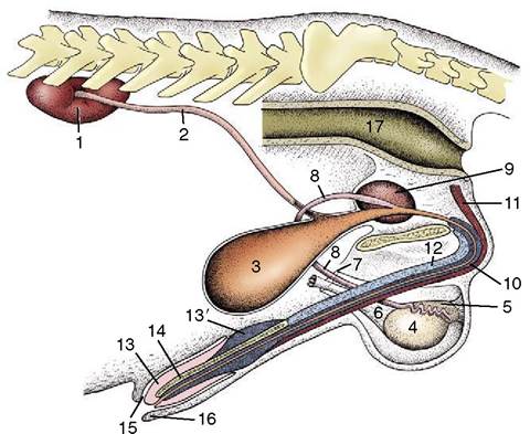
Figure 5-1 The urinary and male reproductive organs (dog). 1, Right kidney; 2, ureter; 3, bladder; 4, testis; 5, epididymis; 6, spermatic cord; 7, vaginal ring; 8, deferent duct; 9, prostate; 10, corpus spongiosum (spongy body); 11, retractor penis; 12, corpus cavernosum (cavernous body); 13, glans penis; 13', bulb of glans; 14, os penis; 15, preputial cavity; 16, prepuce; 17, rectum.
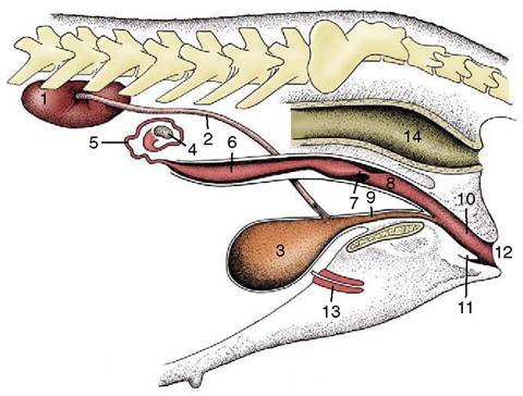
Figure 5-2 The urinary and female reproductive organs (bitch).
1, Right kidney; 2, ureter; 3, bladder; 4, ovary; 5, uterine tube; 6, uterine horn; 7, cervix; 8, vagina; 9, urethra; 10, vestibule; 11, clitoris; 12, vulva; 13, vaginal process; 14, rectum.eventually reaches the cloacal membrane, which is thus divided into dorsal (anal) and ventral (urogenital) parts (Figure 5-5/9). The fusion site corresponds to the perineal body. When the anal membrane breaks down, the dorsal passage becomes a continuous rectoanal canal. A similar rupture of the urogenital membrane
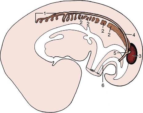
Figure 5-3 Differentiation of the intermediate mesoderm. 1, Pronephros; 2, mesonephros, segmented cranially but continuous caudally; 3, metanephros; 4, pronephric (later mesonephric) duct; 5, ureteric bud; 6, urachus.
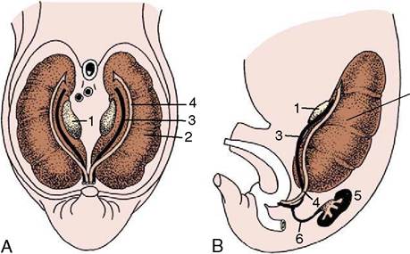
Figure 5-4 Ventral (A) and lateral (B) views of the abdominal roof in a pig embryo of 2.5 cm. The pronephric duct drains the mesonephros and is now more aptly termed the mesonephric duct. 1, Developing gonad; 2, mesonephros; 3, mesonephric duct; 4, paramesonephric duct; 5, metanephros; 6, ureter.
provides the ventral passage with a separate opening to the surface of the body. This urogenital passage differentiates into a cranial part, the future bladder and allantois, and a caudal part from which the urethra is formed.
The bladder then appears as a widening that is continued cranially by the allantoic duct and caudally by an undilated urethra. The allantoic duct or urachus (Figure 5-3/6) can be followed through the umbilical opening to an extraembryonic expansion (the allantois) in which urine accumulates and which is discarded at birth. The part of the duct within the fetus then shrivels
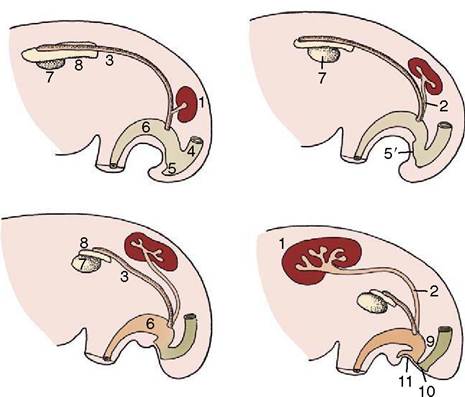
Figure 5-5 The development of the metanephros from two primordia (metanephric cord and ureteric bud).
Note the gradual regression of the mesonephros. 1, Metanephros; 2, ureteric bud (future ureter); 3, mesonephric (deferent) duct; 4, rectum; 5, cloaca; 5, cloacal membrane; 6, urogenital sinus; 7, gonad; 8, remnant of mesonephros (future epididymis); 9, urorectal septum; 10, anal membrane; 11, urogenital membrane.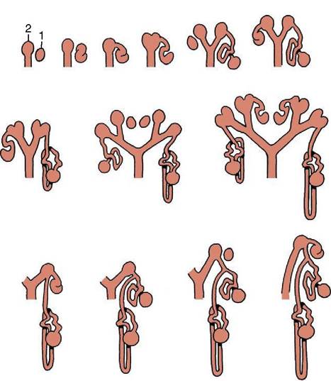
Figure 5-6 This series of schematic drawings depicts the connections between developing nephrons (1) and branches (2) of the ureteric bud. Note the dichotomous division of the drainage system (ureteric bud).
and is finally represented only by the cicatrix or scar on the apex of the bladder. The caudal part of the primordium is transformed into the urethra—the entire urethra in the female but only the short pelvic urethra in the male (in which the penile urethra develops with the genital system). The definitive positions of the openings of the mesonephric and metanephric ducts result from the incorporation of their lower ends within the larger passage. The rearrangement brings the opening of the metanephric duct (ureter) into the bladder, while that of the mesonephric duct (deferent duct) becomes situated more caudally within the urogenital sinus (see Figure 5-5). In this process the mesoderm of the mesonephric duct provides the epithelium of the dorsal trigonal region (p. 183) of the bladder, while the epithelium of the remaining part is provided by hindgut endoderm. The outer layers of the bladder wall differentiate from local mesoderm.