Diagnostics of echinococcosis in dogs
6.1 Clinical diagnosis
The identification of dogs infested with E. granulosus is extremely important for epidemiological studies and monitoring of this zoonosis in control programs [25].
Compared to other gastrointestinal infections of parasitic origin in dogs, echinococcosis is difficult to detect, even if the parasite load is high, premortem diagnosis appears to be very difficult. Recently, there has been considerable progress in research and development of immunological diagnosis for canine echinococcosis. The coproantigen test, in particular, can be considered to be of good sensitivity to reflect a common infestation and can therefore replace purging with arecoline (Table 2).6.2 Parasitological diagnosis
The diagnosis of cystic echinococcosis in ante-mortem dogs can be made using several techniques:
6.2.1 Coproscopy
Coproscopy consists of looking for the eggs or proglottis of the adult worm in the feces of the final host. Eggs can be detected in fecal samples by the flotation technique or on the perianal skin by attaching a transparent adhesive paper to the skin and examining it under a magnifying glass under a microscope. However, this microscopic detection of Echinococcus granulosus eggs is not recommended because of the morphological nature of the egg which is similar in all taenia species. In addition, the removal of eggs is often irregular. However, proglottis from Echinococcus granulosus released spontaneously by dogs and detected on the surface of fecal samples can provide a good diagnosis of parasitosis in dogs [9].
6.2.2 Arecoline purging
Arecoline purging is the standard diagnostic method used for years in the detection of Echinococcus granulosus worms in dogs is purging the intestinal contents
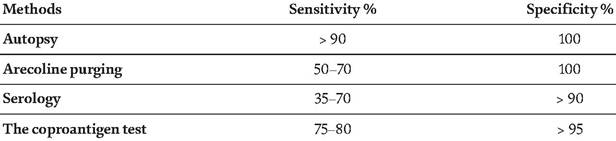
Table 2.
Comparison of the sensitivity and specificity of different diagnostic methods for canine echinococcosis [12].
of the host with hydrobromide arecoline. Arecoline is a parasympathomimetic molecule which has an effect on the smooth muscles of the small intestine and at the same time paralyzes adult worms. The purgation removes the paralyzed worms with the feces, which must be inspected afterwards [9].
The advantage of this technique is the high specificity, which can reach 100%. On the other hand, the sensitivity does not even reach 50% if it is only used once. This test is contraindicated in pregnant dogs, older dogs and puppies. Arecoline should be administered orally at a dose of 4 mg/kg BW. This dose should be carefully calculated since severe undesirable secondary effects may occur [7].
5.1.1 Necropsy diagnosis: Sedimentation and counting
After cutting the intestine into several sections, these must be placed in metal trays, opened with scissors and finally immersed in physiological saline solution. The worms adhering to the mucous membrane are then counted using a magnifying glass or binocular microscope. The disadvantage of this method is that small worms can escape detection [9].
5.2 Immunological diagnosis by detection of Coproantigens
This technique consists of searching for one of two types of antigens, either antigens extracted raw somatically from the worm or excretory-secretory antigens from the protoscolex in the feces of the host using double sandwich ELISA kits [26].
Positive ELISA results can be collected even in the prepatent period, starting on day 5 post-infestation. The values begin to decrease to negative values 2-4 days after the elimination of Echinococcus granulosus worms by treatment with praziquantel. The results of studies using this technique have shown that ELISA values correlate positively with the amount of worms present in the intestine, and that antigen levels are correlated with the amount of worms present in the intestine. This technique has been shown to be important with a sensitivity of 99% and a specificity of 97% [26].
Fecal samples can be taken directly from the ground or rectum and can be kept cold (-20°C) for up to 6 months. The test can be used for the identification of infected cases in control program, including pregnant dogs, older dogs and puppies. Three ELISA kits are commercially available today [27].
5.2.1 Serum antibody detection
Specific serum antibodies (IgG, IgA and IgE) can be detected in the serum of dogs infected with Echinococcus granulosus using antigenic preparations from the protoscolexes in ELISA kits. These antibodies can be detected 2-3 weeks post-infestation. One study suggests that eggs released in the small intestine of the final host, after proglottis apolysis, can penetrate the intestinal barrier and cause immunological stimulation in the host [27].
The ELISA kits available have low sensitivity and highly variable specificity. However, a new kit using a newly derived recombinant antigen from the protoscolex showed 100% specificity, but the sensitivity is not comparable to that of older kits. The use of ELISA kits for the detection of serum antibodies is still questionable because of their low sensitivity, the persistence of antibodies in serum after worm removal and the lack of correlation with infestation pressure [12, 26, 27].
If a seropositive test has been detected but the result is negative for the coproantigens, this is an indication of possible recent exposure [28].
5.3 Molecular biology diagnosis
Parasite DNA can be obtained from eggs, proglottis or worm cells and can be detected in feces after PCR amplification. However, no copro-PCR is currently available for the detection of all strains of Echinococcus granulosus; PCR primers for G1, G5 and combined G6/7 strains have been developed. This technique, due to its high cost, is only used for confirmation on positive samples in areas where the prevalence of cystic echinococcosis is low [29].
6.