DIGESTIVE SECRETIONS
1. What are the two buffers of ruminant saliva and which one of them supports bacterial growth?
2. Is salivary amylase an important component of domestic animal saliva?
3. What is the difference in saliva as related to that resulting from parasympathetic flow and sympathetic flow?
4.
In addition to mucus, what are the gastric secretions?5. What is the relationship of pepsinogen to pepsin?
6. What is the function of pepsin and what is the pH range for its optimal activity?
7. Why would the pH of blood increase (become more alkaline) after ingestion of food? Where is the situation reversed?
8. What is the effect of the barrier presented by tight junctions between gastric cell epithelium?
9. What is rennin and why is it important for young ruminants?
0. What are three substances that stimulate gastric secretions?
11. What are the factors that inhibit gastric secretions?
2. Contrast the pancreatic flowrates between the horse and the dog. Why is there a difference?
3. How is trypsinogen activated? Where does this occur? What activates the other proenzymes?
4. What stimulates the secretion of secretin and cholecystokinin? What is the effect of their secretion?
L5. What is bile? Are bile salts a component of bile? What is meant by recirculation of bile salts? What is the relationship of bile salts to cholesterol? What are gallbladder stones (gallstones)?
6. What controls the contraction of the gallbladder and relaxation of the sphincter of Oddi?
7. Is bicarbonate from the liver (biliary bicarbonate) an important source of bicarbonate for the intestines of some species?
8. What substances from bile emulsify fats?
9. Study Figure 12-35 for a summary of gastrointestinal hormones and association with gastric, pancreatic, and biliary secretions.
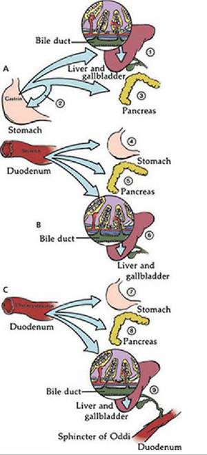
■ FIGURE 12-35 The major mammalian gastrointestinal hormones and their association with gastric, pancreatic, and biliary secretions.
A. Gastrin: 1, stimulates secretion of HCO3- and H2O from bile duct epithelium; 2,,stimulates HCl and pepsinogen secretion; 3, stimulates secretion of pancreatic enzymes. B. Secretin: 4, inhibits HCl secretion and stimulates pepsinogen secretion;.5, stimulates secretion of HCO3- and H2O from pancreas; 6, stimulates secretion of HCO3- and H2O from bile duct epithelium. C. Cholecystokinin: 7, inhibits HCl secretion; 8, stimulates secretion of pancreatic enzymes; 9, stimulates contraction of gallbladder and relaxation of sphincter of Oddi, and stimulates secretion of HCO3- and H2O by bile duct epithelium.In addition to the secretions of the salivary glands, pancreas, and liver, there are those supplied by the many glands of the stomach and intestines that include mucus, hormones, and digestive enzymes. All of these secretions assist the degradation of dietary substances to forms that can be absorbed.
Saliva
In all animal species, salivary secretions facilitate mastication and deglutition because of their watery nature and the lubrication that is provided. The volume of the salivary secretion varies, but is greatest in herbivorous animals. In addition to its lubrication function, saliva increases the potential for evaporation and cooling for panting animals. Saliva has an additional important function in ruminants, in which large volumes of buffered fluid are needed to support microbial fermentation in the rumen and to neutralize the large amounts of acids that are produced as a result of fermentation. To meet the buffering demand, ruminant saliva contains bicarbonate and phosphate buffers. Phosphates are particularly supportive of bacterial growth. In ruminants, salivary secretion is continuous, but the flow of saliva varies with activity and increases with feeding and rumination. Saliva has important antifoaming characteristics and might play a role in reducing the foaming tendency of certain diets.
Consequently, increased salivary flow during eating might help to prevent dietary bloat. In cattle, about 80% of the water entering the stomach is provided by salivary flow derived from extracellular fluid. The need for reabsorption of the water from the large intestine is obvious (see the previous subsection, Intestinal Transport of Electrolytes and Water).The major digestive enzyme produced by the salivary glands is amylase. Among the domestic animals, amylase is most abundant in the saliva of pigs. In contrast, the amount of amylase in human saliva is 100 times that present in pigs.
In addition to the spontaneous secretion of saliva from certain glands in some species (parotid glands in ruminants), secretion is controlled by the autonomic nervous system. Parasympathetic stimulation increases salivary flow that is low in protein (more watery). Sympathetic stimulation, however, has less effect on the flowrate, but increases the amount of protein and mucin and renders saliva more tenacious. The increase in flowrate is brought about by central stimulation from the salivary center and by the mechanical stimulation of receptors in the mouth and stomach. The central component is sometimes referred to as the psychic component (e.g., when an animal salivates in anticipation of food).
Gastric Secretions
In addition to mucus, which is usually secreted throughout the length of the digestive tract, the stomach secretes pepsinogen, HCl, and gastrin. Pepsinogen and HCl are secreted into the lumen of the stomach and gastrin (a hormone) is secreted into the blood. Specific glandular regions are identified within the stomach; their extent varies among species (see Figure 12-7). Generally, the cardiac region secretes only mucus. A variable amount of surface (depending on species) around the cardia has epithelium similar to that of the skin (stratified squamous). This area serves a protective function in the same sense that mucus protects other parts of the digestive tract.
The fundic gland region secretes HCl and pepsinogen (HCl by parietal cells and pepsinogen by neck chief cells) and the pyloric gland region secretes mucus and gastrin. The intrinsic factor is a mucoprotein secreted by the gastric mucosa. It interacts with vitamin B12 to form a complex that binds to receptors in the ileum to facilitate vitamin B12 absorption. The secretion of the intrinsic factor is correlated closely with H+ secretion and it is also secreted by the parietal cells.HCl and pepsinogen initiate the digestion of protein. Pepsinogen is a precursor of pepsin, a proteolytic enzyme. Conversion of the precursor to its active form in the lumen prevents proteolytic digestion of the producing cell. The conversion of pepsinogen to pepsin occurs in the lumen under the influence of HCl and begins at about pH 5. Optimal activity of pepsin occurs at pH 1.8 to 3.5 and initiates gastric protein digestion.
Hydrogen ion is formed in the cell from CO2 according to the hydration reaction^- (- to right of 3 )"? amznid=21960>

H+ is secreted into the stomach lumen and HCO3- is secreted into the blood in exchange for Cl-. The chloride ion is subsequently secreted into the stomach lumen with H+ (Figure 12-33). The increase in plasma bicarbonate concentration that occurs after a meal is known as the alkaline tide, in which the blood pH increases. It is a transient situation that lasts until the pancreas becomes active in secreting HCO3-. An amount equivalent to the amount of HCO3- that entered the blood from the gastric parietal cells is returned to the duodenum by the pancreatic cells.
Because of the high H+ concentration in the stomach, a barrier exists to prevent diffusion of H+ back to the blood. The tight junction between cells is extremely effective and even prevents diffusion of H2O through the epithelium.
This is why highly hypertonic solutions can enter the duodenum because they are not diluted by the diffusion of water into the stomach.In addition to the gastric secretions mentioned above, the young ruminant secretes an enzyme called rennin. This enzyme is a milk-coagulating enzyme; in the presence of Ca2+ it forms a coagulum from milk. This coagulum delays the passage of milk so that more protein digestion occurs in the stomach. The offspring of other animals do not secrete rennin; it is thought that HCl accomplishes the needed coagulation in these animals. The need for rennin in ruminants might relate to their proportionately larger intake of milk at a single nursing, which is not observed for other animals.
Gastric acid secretion is stimulated by acetylcholine, gastrin, and histamine. Acetylcholine is the parasympathetic secretion; it acts directly on the parietal cells to secrete HCl and on the pyloric gland cells to secrete gastrin. Gastrin in turn stimulates HCl and pepsinogen secretion. Chemical releasers of gastrin are digested proteins and amino acids in the stomach. Histamine is an amino
acid derivative present in most body tissues. It is believed that the local gastric mucosal histamine stimulates HCl secretion by potentiating the action of gastrin or by direct stimulation.
Inhibition of gastric acid secretion occurs when the pH of the gastric contents decreases to pH 2 or lower. The acid acts directly on the pyloric gland gastrin cells to inhibit further secretion. Inhibition to gastric acid secretion also originates from the intestine in response to acidic, fatty, and hypertonic solutions entering the duodenum from the stomach, which effectively inhibit gastric emptying. The inhibition is mediated by neural or hormonal mechanisms. The neural mechanism provides inhibitory neurons that synapse with the parasympathetic fibers going to the pyloric gland gastrin cells inhibiting their secretion. The hormonal response is provided by secretin and cholecystokinin (CCK).
CCK occupies the site on the parietal cells that gastrin would have occupied, thus preventing gastrin stimulation of HCl secretion. Secretin works to correct low pH in the duodenum by increasing the production of alkaline secretions that neutralize or buffer the acid.The factors that regulate gastric secretions can be summarized as follows:
1. Stimulation
a. Acetylcholine
b. Gastrin
c. Histamine
2. Inhibition
a. Within stomach: decrease of pH to 2
b. From duodenum: presence of acidic, fatty, and hypertonic solutions
i. Neural mechanism - inhibitory neurons to parasympathetic fibers that then inhibit the secretion of gastrin
ii. Hormonal mechanism - secretion of CCK and secretin: CCK prevents gastrin stimulation of HCl secretion; secretin increases production of alkaline secretions
Pancreatic Secretions
Only the exocrine secretions (HCO3- and digestive enzymes or precursors) of the pancreas are involved in the digestive process. The secretion of HCO3- is needed to neutralize the HCl concentration of the stomach contents that enter the duodenum and also for neutralization of acids produced from fermentation in the large intestine. Enzymes and enzyme precursors are needed for digestion in the intestinal lumen so that the products of degradation can be absorbed. These secretions are somewhat more unique in omnivores and nonruminant herbivores. In these animals, a large volume of buffered fluid is needed for the microbial digestion that occurs in the cecum and colon. The digestive enzymes provide for small intestine digestion and the larger volume of fluid and HCO3- serves a function similar to that of saliva in the ruminant. In the horse, the rate of enzyme secretion is low in comparison with that of other species. This might occur because a greater proportion of the horse’s ingested food is of a type that requires microbial digestion beyond the small intestine.
There is a continuous flow of pancreatic fluid in the horse, even under basal (nonfeeding) conditions. This ensures that an adequate volume of buffered fluid (containing HCO3-) is present for the continuous fermentation in the cecum and colon. The rate can be increased under stimulation. In contrast, the dog might have almost no fluid flow from the pancreas under basal conditions, but high rates of flow are produced under stimulation. This pattern is appropriate because the dog eats less frequently and because little fermentation occurs in the large intestine and a large volume of buffered fluid is not needed.
The pancreas secretes all of the enzymes and enzyme precursors (proenzymes) necessary for the digestion of proteins, fats, and carbohydrates. The proteases are secreted in proenzyme form and include trypsinogen, chymotrypsinogen, elastase, and carboxypeptidases A and B. Trypsinogen is activated by enterokinase (present in the intestinal epithelium) to form trypsin only after it reaches the intestinal lumen, and the reaction occurs at the brush border. Trypsin then becomes the activator for the other proenzymes. Digestion of the pancreas is prevented because the proteolytic enzymes are secreted as proenzymes. Spontaneous conversion of trypsinogen to trypsin is prevented in the pancreas by the presence of trypsin inhibitor.
Pancreatic lipase hydrolyzes dietary triglycerides into substances that can then be absorbed. Bile salts are needed to activate pancreatic lipase.
Pancreatic amylase is secreted in its active form. This carbohydrate enzyme hydrolyzes starch to maltose, a disaccharide. No free glucose is formed by pancreatic amylase hydrolysis.
The exocrine secretions of the pancreas are controlled by autonomic nerves as well as by the gastrointestinal hormones gastrin, CCK, and secretin. Parasympathetic stimulation increases the secretion of enzymes and proenzymes, with little secretion of electrolytes and water in most species. Increased water and electrolyte secretion, however, does accompany parasympathetic stimulation in the pig and horse (these animals need large volumes of water and HCO3- for large intestine fermentation). Gastrin that is secreted when the parasympathetics are stimulated can also stimulate the pancreas to release enzymes and proenzymes, whereby the parasympathetic effect on the pancreas is potentiated. Two hormones secreted when the stomach contents enter the intestine are secretin and CCK. Secretin release is stimulated by acid perfusion of the duodenum and causes the pancreas to secrete HCO3-. Secretin was the first hormone discovered, which occurred in 1902, as the result of work by Bayliss and Starling. The hormone CCK is secreted in response to the presence of protein and fat in the duodenum and causes the pancreas to.secrete enzymes and proenzymes. Secretin and CCK are synergistic to each other - that is, the presence of one enhances the effect of the other.
Biliary Secretions
Bile is a greenish-yellow solution of bile salts, bilirubin, cholesterol, lecithin, and electrolytes (Na+, K+, Cl-, and HCO3-). Bile salts are synthesized continuously by hepatic cells, but the quantity needed for digestion far exceeds the rate of production by the liver. Therefore, bile salts are recirculated from the intestine (after being used in the intestine) to the hepatic cells, where they are resecreted (enterohepatic circulation). Because of this reuse of bile salts, an adequate amount is available for efficient digestion. Bile salts are synthesized from cholesterol and, in the process, some cholesterol, as well as bile salts, is secreted into the bile. The bile salts and lecithin form a soluble micelle (colloidal particle) with cholesterol, thereby preventing cholesterol precipitation and gallstone formation. The solubility, however, depends on an alkaline solution, which is provided by HCO3-.
Bile is secreted continuously in all species and can be transported to the gallbladder and stored for later use or transported directly to the intestine. While in the gallbladder, bile can be concentrated by absorption of NaCl or of NaHCO3 and water. The degree of concentration depends on the length of storage. In animals that eat once or twice per day,,bile is highest in concentration, but its, concentration is low in ruminants and the pig because they eat frequently and bile is therefore discharged from the gallbladder frequently. The horse does not have a gallbladder and a large flow of hepatic bile continuously enters the duodenum; it is the only domestic animal without a gallbladder. The opening of the common bile duct into the duodenum is controlled by the sphincter of Oddi. Contraction of the gallbladder and relaxation of the sphincter are controlled by CCK, which is released in response to the presence of lipids and amino acids in the small intestine.
Bile secretion by the liver is primarily stimulated by the amount of bile salts being recirculated. On reaching the liver, the bile salts are absorbed from the hepatic sinusoids (portal circulation) into the hepatic cells and are then resecreted into the bile canaliculi by active transport (Figure 12-34). Cations and water diffuse passively, so that the newly formed bile is iso-osmotic with plasma. Therefore, the larger the amount of bile salts recirculated, the higher is the rate of secretion of bile. Bicarbonate and other electrolytes are secreted by the bile duct epithelium as well (biliary secretion), and their secretion is increased by CCK, secretin, and gastrin. The secretion of biliary bicarbonate is an important source of buffer for the intestine in some species. In the sheep, the rate of biliary secretion of HCO3- is much higher than that of the pancreatic secretion and the liver plays a greater role than the pancreas in neutralizing the H+ in the duodenum.
Fat in the intestine is emulsified (breakdown of fat globules into smaller globules) by the bile salts and by the lecithin present in the bile. This provides a greater surface area for digestion by the luminal lipases (lipid enzymes). Another important function of the bile salts is the removal of the products of lipid digestion (free fatty acids and monoglycerides) from the area of digestion (transport function) so that digestion can continue without recombination to triglycerides. The bile salts accomplish this transport function by forming soluble micelles. In this form the digestion products are moved easily by diffusion to the intestinal epithelium for absorption.
A summary of the major gastrointestinal hormones and their association with gastric, pancreatic, and biliary secretions is presented in Figure 12-35.
■